FIGURE SUMMARY
Title

PAX1 represses canonical Wnt signaling pathway and plays dual roles during endoderm differentiation

Authors
Miao, D., Ren, J., Jia, Y., Jia, Y., Li, Y., Huang, H., Gao, R.
Source
Full text @ Cell Commun. Signal.

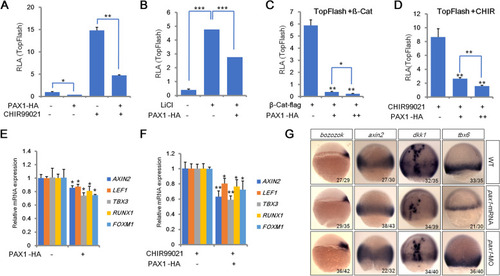
PAX1 plays a negative role in canonical Wnt signaling pathway. A-B TopFlash luciferase reporter assays in HEK293FT cells transfected with an empty vector control or a PAX1 expression plasmid, with or without treatment of GSK3 inhibitor CHIR99021 (CHIR) (A) or LiCl (B). RLA: relative luciferase activity. C-D TopFlash luciferase reporter assays in HEK293FT cells with transfection of an increasing dose of HA-tagged PAX1 and activated Wnt signaling by co-transfection of flag-tagged β-catenin (β-Cat) (C) or treatment with CHIR99021 (D). E-F Quantitative real-time PCR (qRT-PCR) analyses of Wnt target genes performed in HEK293FT cells with or without CHIR99021 treatment. G Expression of Wnt target genes by in situ hybridization in WT (wild type) control, pax1 mRNA or morpholino (MO) injected zebrafish embryos. The frequency of embryos with the indicated patterns is shown in the bottom right corner of each group. Bar graphs and error bars represent mean ± standard deviation (SD) of at least three biologically independent experiments. *P < 0.05; **P < 0.01; ***P < 0.001. This is the same in the following figures unless specifically indicated

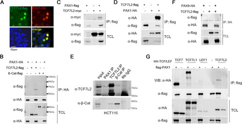
PAX1 interacts with TCF7L2. A Immunofluorescence showed the subcellular localization of TCF7L2 and PAX1 by co-transfection of TCF7L2-flag and PAX1-HA tag fused expression plasmids in HEK293FT cells. DAPI staining revealed the nuclei. Merged image showed that TCF7L2 and PAX1 are co-localized in the nucleus. B-D PAX1 and TCF7L2 or β-catenin (β-Cat) expression plasmids were transfected alone or together into HEK293FT cells and their interaction was determined by co-immunoprecipitation (co-IP) and western blotting. TCL: total cell lysate. E The endogenous interaction of PAX1 and TCF7L2 was tested in HCT116 cells. β-Cat and TCF7L2 IPs were used as positive controls. F PAX9 and TCF7L2 expression plasmids were transfected alone or together into HEK293FT cells and their interaction was determined by co-IP and western blotting. G The interactions between PAX1 and TCF/LEF family members were assessed by co-IP assays and western blotting, with PAX1 and TCFs expression plasmids transfection alone or together into HEK293FT cells

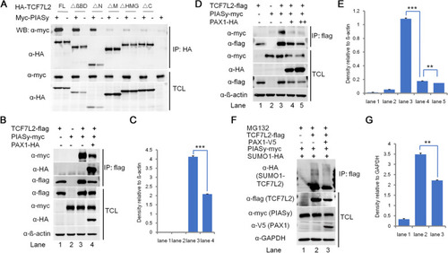
PAX1 inhibits the interaction between TCF7L2 and PIASy, and decreases the SUMOylation level of TCF7L2. A PIASy interacts with the middle, HMG and C-terminal domains of TCF7L2. Different HA-tagged TCF7L2 deletion constructs depicted in Fig. S2A were transiently transfected into HEK293FT cells together with Myc-tagged PIASy. Cell lysates were immunoprecipitated with M2 beads (anti-flag), followed by western blotting using anti-HA for TCF7L2 proteins and anti-Myc for PIASy. B Interactions between TCF7L2 and PIASy were assessed by co-IP and western blotting with empty vector control or PAX1-HA cotransfection. HEK293FT cells were transfected with flag-tagged TCF7L2 together with PIASy-myc and PAX1-HA plasmids. Following co-IP with M2 beads, TCL and IP samples were assayed by western blotting using anti-myc antibody for PIASy and anti-HA antibody for PAX1, respectively. ß-actin was used as loading control. C Densitometry analysis of western blots in (B) (n = 2, mean ± SD. *P < 0.05, **P < 0.01, ***P < 0.001). D PAX1 disturbs the interaction between TCF7L2 and PIASy in a dose-dependent manner. Same as (B) except increasing amount of HA-PAX1 (0 μg, 1 μg, and 2 μg) were transfected into HEK293FT cells together with flag-TCF7L2 and myc-PIASy. When flag-tagged TCF7L2 was immunoprecipitated by M2 beads (anti-flag), the amount of coprecipitated Myc-tagged PIASy was negatively correlated with the dose of cotransfected PAX1. E Densitometry analysis of western blots in (D) (n = 2, mean ± SD. *P < 0.05, **P < 0.01, ***P < 0.001). F Western blotting analysis of SUMOylation level of TCF7L2. HEK293FT cells were cotransfected with plasmids expressing flag-TCF7L2, V5-PAX1, and HA-tagged SUMO1. Cells were treated with proteasome inhibitors MG132 (10 μM) for 6 hours before harvested. GAPDH was used as loading control. G Densitometry analysis of western blots in (F) (n = 2, mean ± SD. *P < 0.05, **P < 0.01, ***P < 0.001)

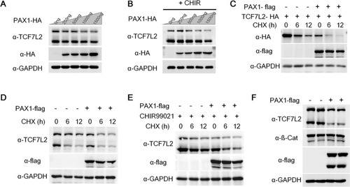
PAX1 reduces TCF7L2 protein stability. A HEK293FT cells were transfected with increasing amount of PAX1-HA (0, 250, 500, 1000, 1500 ng) expression plasmid and endogenous TCF7L2 levels were analyzed by western blotting. Both bands shown in TCF7L2 blot indicate different splicing isoforms of human TCF7L2 expressed in HEK293FT cells. GAPDH was used as loading control. B Same as (A) except cells were treated with 10 μM CHIR99021 (+CHIR) for 24 hours before harvested. C HEK293FT cells were transfected with TCF7L2-HA and empty vector (−) or PAX1-flag plasmid (+) and 2 days later were treated with 100 μg/ml cycloheximide (CHX) for indicated time and harvested. The expression of TCF7L2 (HA), PAX1 (flag), and GAPDH were analyzed by western blotting. D HEK293FT cells were transfected with empty vectors (−) or PAX1-flag plasmids (+) and cells were treated with 100 μg/ml CHX for indicated time before harvested. Endogenous TCF7L2 levels were analyzed by western blotting. GAPDH was used as loading control. E Same as (D) except cells were treated with 10 μM CHIR99021 (CHIR) for 24 hours before harvested. F Western blotting to analyze the protein levels of indicated proteins in WT and PAX1 overexpression (OE) HCT116 cell lines. GAPDH was used as loading control. Both bands detected in the flag blot should be PAX1 isoforms (NCBI database: NP_006183.2 and NP_001244025.1) which contains 534 and 457 amino acids respectively

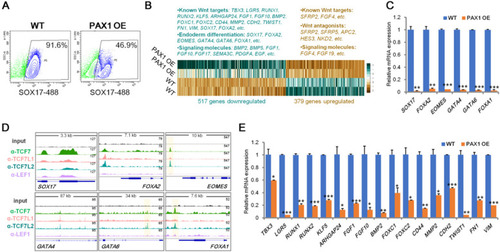
PAX1 ectopic expression leads to impaired definitive endoderm differentiation by inhibiting Wnt signaling. A Flow cytometry analysis of wild-type (WT) control and PAX1 overexpressed (OE) hESC-derived definitive endoderm cells stained for SOX17 (Alexa Fluor 488) demonstrates decreasing of SOX17+ population when PAX1 was ectopically expressed for a representative differentiation (n = 3 independent differentiations). B Heatmap representation of RNA-seq analysis of definitive endoderm cells derived from WT and PAX1 OE hESCs (n = 2; log2 fold change ≤ − 1, ≥1; p-value < 0.05). Differentially expressed known Wnt targets, endoderm markers and signaling molecules were listed above. C qRT-PCR analyses of endoderm marker genes in hESC-derived definitive endoderm cells. D Genome tracks of TCF7, TCF7L1, TCF7L2 and LEF1 ChIP-seq reads analyzed from datasets (GSE182842) on definitive endoderm marker genes in human pluripotent stem cell-derived definitive endoderm cells. E qRT-PCR analyses of known Wnt target genes in hESC-derived definitive endoderm cells

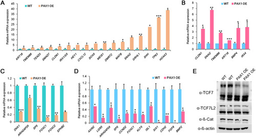
PAX1 represses Wnt signaling pathway in foregut and pharyngeal endoderm cells. A-B qRT-PCR analyses of AFE and PE associated genes in hESC-derived AFE (A) and PE (B) cells. AFE: anterior foregut endoderm; PE: pharyngeal endoderm. PAX1 OE: PAX1 overexpression. C-D qRT-PCR analyses performed on known Wnt target genes in hESC-derived AFE (C) and PE (D) cells. E Western blotting to analyze the protein levels of indicated proteins in WT and PAX1 OE hESC-derived PE cells. ß-actin was used as loading control

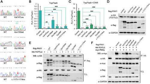
The roles of PAX1 mutants associated with SCID in regulation of Wnt signaling pathway. A Sanger sequencing results of wild type (WT) control and SCID associated PAX1 mutation constructs. B-C TopFlash luciferase reporter assays performed in HEK293FT cells transfected with empty vector control, WT or PAX1 mutation constructs without (B) or with (C) treatment of CHIR99021 (CHIR). * represents significant decrease compared with control, while # represents significant increase compared with PAX1 WT. D HEK293FT cells were transfected with HA-tagged TCF7L2 and empty vector control, PAX1 WT or PAX1 mutation construct (flag-tagged). TCF7L2 levels were analyzed by western blotting with anti-HA antibody. GAPDH was used as loading control. E A series of PAX1 mutation constructs with flag-tag were co-transfected with HA-tagged TCF7L2 in HEK293FT cells. Co-IP and western blotting assays were performed to test the interactions between TCF7L2 and PAX1 proteins. F Interactions between TCF7L2 and PIASy were assessed by co-IP and western blotting with empty vector control, PAX1 WT or PAX1 mutation constructs cotransfection in HEK293FT cells. Co-IP was done with anti-HA antibody. TCL and IP samples were assayed by western blotting using anti-myc antibody for PIASy and anti-flag antibody for PAX1 proteins, respectively. GAPDH was used as loading control

Acknowledgments
This image is the copyrighted work of the attributed author or publisher, and ZFIN has permission only to display this image to its users. Additional permissions should be obtained from the applicable author or publisher of the image. Full text @ Cell Commun. Signal.