FIGURE

Fig. 2

ID
ZDB-FIG-240429-2
Publication
Miao et al., 2024 - PAX1 represses canonical Wnt signaling pathway and plays dual roles during endoderm differentiation
Other Figures
All Figure Page
Back to All Figure Page
Fig. 2

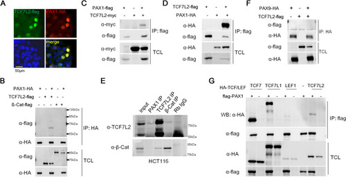
PAX1 interacts with TCF7L2. A Immunofluorescence showed the subcellular localization of TCF7L2 and PAX1 by co-transfection of TCF7L2-flag and PAX1-HA tag fused expression plasmids in HEK293FT cells. DAPI staining revealed the nuclei. Merged image showed that TCF7L2 and PAX1 are co-localized in the nucleus. B-D PAX1 and TCF7L2 or β-catenin (β-Cat) expression plasmids were transfected alone or together into HEK293FT cells and their interaction was determined by co-immunoprecipitation (co-IP) and western blotting. TCL: total cell lysate. E The endogenous interaction of PAX1 and TCF7L2 was tested in HCT116 cells. β-Cat and TCF7L2 IPs were used as positive controls. F PAX9 and TCF7L2 expression plasmids were transfected alone or together into HEK293FT cells and their interaction was determined by co-IP and western blotting. G The interactions between PAX1 and TCF/LEF family members were assessed by co-IP assays and western blotting, with PAX1 and TCFs expression plasmids transfection alone or together into HEK293FT cells

Expression Data

Expression Detail
Antibody Labeling
Phenotype Data

Phenotype Detail
Acknowledgments
This image is the copyrighted work of the attributed author or publisher, and ZFIN has permission only to display this image to its users. Additional permissions should be obtained from the applicable author or publisher of the image. Full text @ Cell Commun. Signal.