FIGURE SUMMARY
Title

Plexin B3 guides axons to cross the midline in vivo

Authors
Liu, Z.Z., Liu, L.Y., Zhu, L.Y., Zhu, J., Luo, J.Y., Wang, Y.F., Xu, H.A.
Source
Full text @ Front. Cell. Neurosci.

Plexin B3 is expressed in retinal ganglion cells when retinal axons are crossing the midline. In situ hybridization was performed on Tg[Isl2b:GFP] transgenic embryos at 34–36 hpf (hour post fertilization), in which the reporter protein GFP is expressed specifically in retinal ganglion cells and can also label retinal axons. (A) Fluorescent in situ hybridization using complementary RNA probe reveals that Plexin B3 is strongly expressed in retina at 34 hpf. (B) Tg[Isl2b:GFP] transgenic labeling demonstrates that Plexin B3 is strongly expressed in retinal ganglion cell layer (square brackets) when pioneer retinal axons are starting to cross the midline and form the chiasm. (C,D) Plexin B3 is expressed in retinal axon ganglion cell layer at 36 hpf when many retinal axons cross the midline. (E,F) A zoomed in and single plane of confocal image demonstrates that Plexin B3 is expressed in retinal ganglion cell layer. All images are ventral view, rostral to the top. Scale bar, 100 μm.

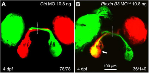
Knocking down Plexin B3 induces retinal axons to misproject into the ipsilateral tectum. (A) In control MO treated embryos, all retinal axons cross the midline and project to the contralateral tectum. (B) In some of the MOE4 treated embryos (36 out of 140 eyes), retinal axons fail to cross the midline at the chiasm and misproject to the ipsilateral tectum (arrow). Images are dorsal view, rostral to the top. Scale bar, 100 μm.

PHENOTYPE:
Fish:
Knockdown Reagent:
Observed In:
Stage: Day 4

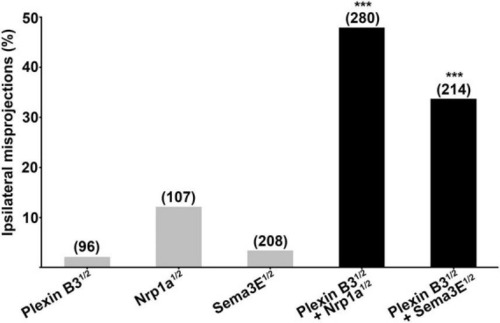
Plexin B3 synergizes genetically with Neuropilin1 and Sema3E in retinal axon guidance. Half doses of Plexin B3, Nrp1a or Sema3E MO results in low proportion of ipsilateral misprojections of retinal axons. Combining half doses of Plexin B3 MO with Nrp1a or Sema3E MO causes dramatic increase of ipsilateral misprojections, which is much more than the simple sum up of the individual half doses of MOs. The combo results strongly indicate that Plexin B3 genetically interact with Nrp1a and Sema3E and might function as receptor-ligand in axon guidance in vivo. Stars above the columns indicate that the proportion of eyes with ipsilateral misprojections induced by the combination of half doses is significantly higher than would be expected by assuming that the effects of half doses add together independently. ***p < 0.001, Fisher’s exact test.

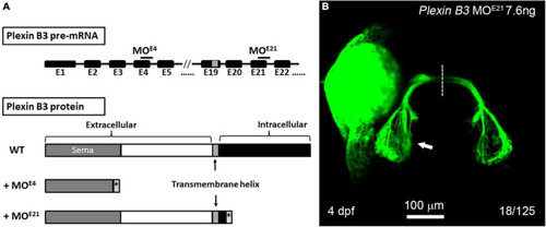
The intracellular domain of Plexin B3 is required for signaling transduction in retinal axon guidance. (A) Two morpholinos were designed to interfere with the splicing of exon 4 (E4) and exon 21 (E21) of Plexin B3 pre-mRNA. Sequencing the mis-spliced mRNA revealed that MOE4 results in a truncated protein remaining only part of extracellular part and MOE21 results in a truncated protein missing the majority of intracellular domains. (B) Plexin B3 MOE21 treatment causing misprojection of retinal axons into the ipsilateral tectum (arrow), similar as the effect of MOE4. The image is dorsal view, rostral to the top. Scale bar, 100 μm.

PHENOTYPE:
Fish:
Knockdown Reagent:
Observed In:
Stage: Day 4

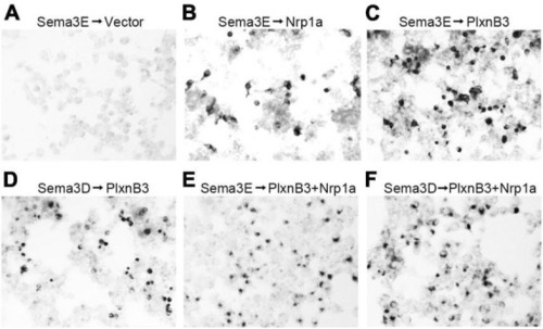
Sema3E binds to Plexin B3 in vitro. The binding assay was performed using type 3 Semaphorins tagged with alkaline phosphatase (AP-Sema3) according to He et al. (2019). (A) Supernatant containing AP-tagged Sema3E was applied to HEK293 cells transfected with pAG3 empty vector. No obvious binding of Sema3E to the cells was detected. (B) AP-tagged Sema3E binds to cells expressing Nrp1a. (C,D) Sema3E or Sema3D binds to Plexin B3. (E,F) Sema3E or Sema3D binds to the cells co-transfected with Plexin B3 and Nrp1a.

Acknowledgments
This image is the copyrighted work of the attributed author or publisher, and ZFIN has permission only to display this image to its users. Additional permissions should be obtained from the applicable author or publisher of the image. Full text @ Front. Cell. Neurosci.