FIGURE SUMMARY
Title

Maternal vgll4a regulates zebrafish epiboly through Yap1 activity

Authors
Camacho-Macorra, C., Tabanera, N., Sánchez-Bustamante, E., Bovolenta, P., Cardozo, M.J.
Source
Full text @ Front Cell Dev Biol

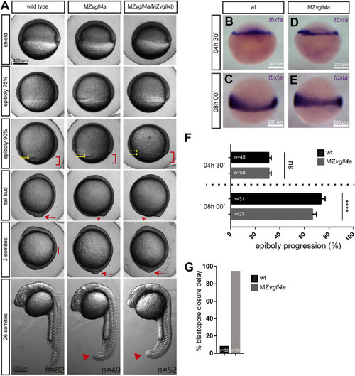
Epiboly progression in MZvgll4a embryos is delayed. (A) Bright field images of wt, MZvgll4a and MZvgll4a;MZvgll4b embryos at different developmental stages as indicated in the panels for wt embryos. Note the delayed epiboly (red brackets) and blastopore closure (red asterisks) as well as the increased distance between the EVL and DEL margins (yellow arrows) in MZvgll4a and MZvgll4a;MZvgll4b embryos as compared to wt. Blastopore closure (red arrow) in mutants occurs when wt embryos have initiated somitogenesis (red dotted line). At the 26 somites’ stage, the tail of mutants is still not fully elongated (red arrowheads). (B–E) Margin cells in wt (B,C) and MZvgll4a(D,E) embryos stained by in situ hybridization of tbtxa at 4.5hpf (B,D) and 8hpf (C,E). (F) Quantification of epiboly progression of wt and MZvgll4a embryos stained by in situ hybridization of tbtxa at 4.5hpf and 8hpf. T-test, p < 0.0001. (G) Quantification of wt and MZvgll4a embryos that do not reach blastopore closure at 10hpf. Fisher’s exact test, p < 0.0001.

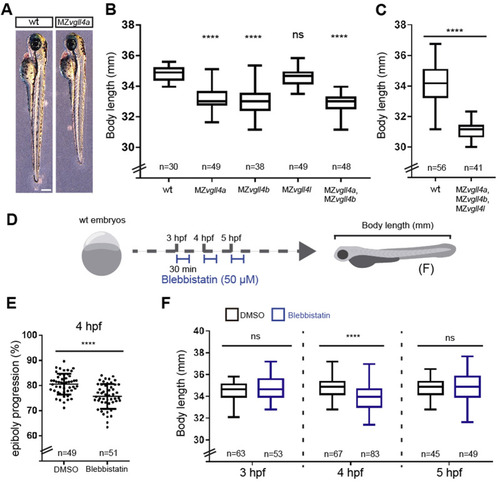
Body length is affected in MZvgll4 larvae which could be related to epiboly progression delay. (A) Lateral views of wt and MZvgll4a larvae at 3dpf (left) and graph (B) showing the quantification of the body length from wt, MZvgll4a, MZvgll4b, MZvgll4l and MZvgll4a;MZvgll4b larvae at 3dpf. Note that MZvgll4a, MZvgll4b and MZvgll4a;MZvgll4b but not MZvgll4l mutants are shorter that the wt. Mann Whitney test, p < 0.0001. (C) The body length of MZvgll4a;MZvgll4b;MZvgll4l triple mutant larvae are also shorter than wt at 3dpf. Kruskal–Wallis test, p < 0.0001. (D) Schematic representation of the experimental design in (F). (E) The blebbistatin treatment at 4hpf during 30 min, delays epiboly progression in wt embryos (F) The treatment of wt embryos with blebbistatin at 4hpf during 30 min affects the body length of the larvae at 3dpf. Mann Whitney test, p < 0.0001. But not at 3hpf nor 5 hpf. Mann Whitney test, p = 0.2545 and p = 0.4396 respectively. T-test, p < 0.0001. Ns, not significant. ****p < 0.0001.

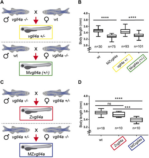
Zygotic vgll4a function is dispensable for timely embryonic growth. (A,C) Schematic representation of the mating strategy used to obtain embryos of the desired genotypes with or without vgll4a maternal contribution. (B,D) Box plots of the body length from 3dpf larvae of the indicated genotypes. Note the decreased body length only in embryos with absent or haplo-insufficient maternal vgll4a contribution as compared to wt. The number of analyzed embryos is indicated below each plot. Data in B were analyzed with Kruskal–Wallis test, p < 0.0001, whereas those in D with One-Way ANOVA. ns, not significant. ***p < 0.001. ****p < 0.0001.

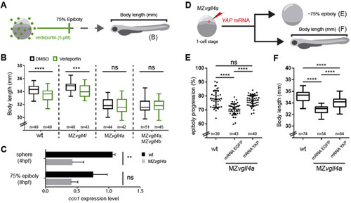
Vgll4a acts upstream of yap activity to promote embryonic growth. (A) Schematic representation of the experimental design in (B). (B) Box plots of the body length from wt, MZvgll4l, MZvgll4a and MZvgll4a;MZvgll4b embryos grown in the presence of verteporfin or DMSO. Note that the drug has no effect on the already reduced body length of MZvgll4a and MZvgll4a;MZvgll4b larvae but reduces that of wt and MZvgll4l larvae. T-test. wt, p < 0.0001; MZvgll4l, p = 0.0003; MZvgll4a, p = 0.5769 and MZvgll4a;MZvgll4b, p = 0.9744. (C) The graphs show the expression level of the Yap-TEAD transcriptional target ccn1 at sphere and 75% epiboly stage in wt and MZvgll4a embryos as determined by qRT-PCR analysis. Note the significant reduction of ccn1 expression in the mutants. T-test. At 4hpf, p = 0.0042 and at 8hpf, p = 0.0746. (D) Schematic representation of the experimental design of data reported in (E,F). (E) YAP mRNA injection rescues epiboly progression in MZvgll4a embryos to an extend similar to that of wt embryos whereas eGFP mRNA has no effect. Kruskal–Wallis test, p < 0.0001. wt vs. MZvgll4a mRNA YAP injected, p > 0.9999. (F) MZvgll4a embryos injected with YAP mRNA display a larvae body length larger than MZvgll4a embryos injected with control mRNA. One-Way ANOVA, p < 0.0001. Ns, not significant. **p < 0.01. ***p < 0.001. ****p < 0.0001.

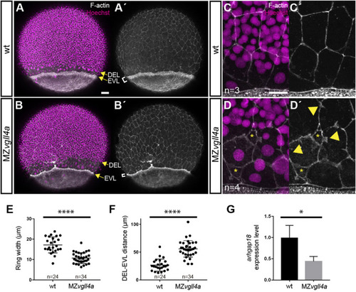
Maternal vgll4a is required for proper organization of the actomyosin ring. (A-D′) Confocal images of wt and MZvgll4a embryos at 75% epiboly stage (lateral views) stained with phalloidin and Hoechst to visualize F-actin and nuclei, respectively. Note that the actomyosin ring in MZvgll4a embryos is thinner (white brackets in A′, B′) and rather separated from the DEL margin (yellow arrows in A, B) as compared to wt embryos. Note also that F-actin distribution of EVL cells in MZvgll4a embryos is ruffled and disorganized (yellow arrowheads in D′) in contrast to the well aligned distribution in wt embryos. Asterisk in D, D′ indicates loss of cell-cell contacts. (E,F) Quantification of the actomyosin ring width and DEL-EVL distance in wt (n = 24) and MZvgll4a (n = 34) embryos. Mann-Whitney test, p < 0.0001). (G) The graphs show the expression level of arhgap18 transcripts in MZvgll4a and wt embryos at 75% epiboly stage. t-test, p = 0.041, as determined by qRT-PCR analysis. *, p < 0.05. ****, p < 0.0001. Scale Bars, a-b´ 50µm, c-d´, 20 µm.

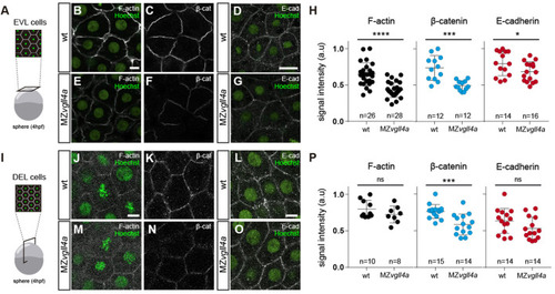
Maternal vgll4a is required for plasma membrane localization of the E-cadherin/β-catenin complex. (A,I) Schematic representation of the different imaging strategies. (B-G; J-O) Confocal images of F-actin (B, E, J, M), β-catenin (C, F, K, N) and E-cadherin (D, G, L, O) distribution in EVL (B–G) and DEL (J–O) cells in wt and MZvgll4a embryos at sphere stage. Embryos were counterstained with Hoechst (nuclei, green). (H,P) The graphs depict the fluorescent signal intensity (in arbitrary units, a. u) for F-actin, β-catenin and E-cadherin in EVL (H) or DEL (P) cells of wt and MZvgll4a embryos. Mann Whitney test. (H) β-catenin, p = 0.0001, E-cadherin; p = 0.0425. (P) F-actin, p = 0.3154; β-catenin, p = 0.0006 and E-cadherin, p = 0.1599. Ns, not significant, *p < 0.05; ***p < 0.001; ****p < 0.0001. Scale bar, 10 µm.

Proposed model for maternal vgll4a contribution to zebrafish epiboly progression. Maternal vgll4a promotes plasma membrane localization of the E-cadherin/β-catenin complex in the amount required for an adequate cohesion among blastomeres. This cohesion threshold allows tissue mechano-sensing and thus yap1-dependent mechano-transduction. Signal transduction impacts on the organization and function of the actomyosin ring, thereby promoting timely epiboly progression. E-cad, E-cadherin; β-cat, β-catenin; F-act, F-actin.

Acknowledgments
This image is the copyrighted work of the attributed author or publisher, and ZFIN has permission only to display this image to its users. Additional permissions should be obtained from the applicable author or publisher of the image. Full text @ Front Cell Dev Biol