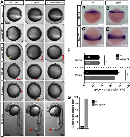
Epiboly progression in MZvgll4a embryos is delayed. (A) Bright field images of wt, MZvgll4a and MZvgll4a;MZvgll4b embryos at different developmental stages as indicated in the panels for wt embryos. Note the delayed epiboly (red brackets) and blastopore closure (red asterisks) as well as the increased distance between the EVL and DEL margins (yellow arrows) in MZvgll4a and MZvgll4a;MZvgll4b embryos as compared to wt. Blastopore closure (red arrow) in mutants occurs when wt embryos have initiated somitogenesis (red dotted line). At the 26 somites’ stage, the tail of mutants is still not fully elongated (red arrowheads). (B–E) Margin cells in wt (B,C) and MZvgll4a(D,E) embryos stained by in situ hybridization of tbtxa at 4.5hpf (B,D) and 8hpf (C,E). (F) Quantification of epiboly progression of wt and MZvgll4a embryos stained by in situ hybridization of tbtxa at 4.5hpf and 8hpf. T-test, p < 0.0001. (G) Quantification of wt and MZvgll4a embryos that do not reach blastopore closure at 10hpf. Fisher’s exact test, p < 0.0001.
|

