FIGURE SUMMARY
Title

Color-Flu Fluorescent Reporter Influenza A Viruses Allow for In Vivo Studies of Innate Immune Function in Zebrafish

Authors
Soos, B.L., Ballinger, A., Weinstein, M., Foreman, H., Grampone, J., Weafer, S., Aylesworth, C., King, B.L.
Source
Full text @ Viruses

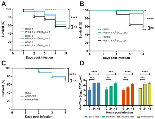
Characterization of PR8 and Color-flu systemically infected AB zebrafish. (A) Decreased survival of AB zebrafish systemically infected with three different lots of PR8 IAV at 2 dpf compared to vehicle (HBSS) controls (p < 0.0001 for each lot comparison). Survival rates of PR8-infected embryos were not significantly different between lots (p = 0.0603). (B) Decreased survival of AB zebrafish systemically infected with two different lots of PR8 IAV at 3 dpf compared to vehicle controls (p < 0.0001 for each lot comparison). Survival rates of PR8-infected embryos were not significantly different between lots (p = 0.1442). (C) Decreased survival of AB zebrafish systemically infected with eCFP-PR8 or Venus-PR8 (3.2 × 10² TCID50/mL) compared to vehicle controls (p < 0.0001 for each comparison). Survival rates of eCPF-PR8- or Venus-PR8-infected zebrafish were not significantly different strains (p = 0.5238). (D) Increased TCID50 viral titer in Color-flu systemically infected AB zebrafish at 24 and 48 hpi compared to 0 hpi. eCFP-PR8-, mCherry-PR8-, and Venus-PR8-infected zebrafish had increased viral titers at 24 and 48 hpi compared to 0 hpi (adjusted p-values = 0.0001 and <0.0001, respectively, for eCFP-PR8; 0.0019 and 0.0001, respectively, for eGFP-PR8; 0.0047 and <0.0001, respectively, for mCherry-PR8; and 0.0003 and <0.0001, respectively, for Venus-PR8). Survival studies were conducted with four independent experiments (n = 4) and 50 larvae per sample group. TCID50 assays were carried out with three independent experiments (n = 3) and 25 larvae per group for each time point. Not significant (ns), p > 0.05; ** p < 0.01; *** p < 0.001; **** p < 0.0001.

Characterization of PR8 and Color-flu systemic infection across zebrafish lines. (A) Decreased survival was greater in casper than AB or EK lines with 2 dpf systemic PR8 infection. PR8-infected zebrafish had a decreased survival rate compared to vehicle (HBSS) controls (p < 0.0001 for all lines). Casper zebrafish had a lower survival rate than EK (p = 0.0024) and AB (p = 0.0055) according to the log-rank Mantel–Cox test. (B) Decreased survival rates were observed in AB, EK, and casper with 3 dpf PR8 infection compared to controls (p < 0.0001 for all lines). No significant (ns) difference in survival rate was detected between PR8-infected AB, EK, and casper larvae. (C) Increased TCID50 viral titer in 2 dpf PR8-infected AB, EK, and casper zebrafish at 24, 48, and 72 hpi compared to 0 hpi (adj. p-value < 0.001 for all comparisons except for AB 24 hpi (adj. p-value = 0.0006) and AB 96 hpi (adj. p-value = 0.0003)). (D) Increased TCID50 viral titer in 3 dpf PR8-infected AB, EK, and casper zebrafish at 72 hpi compared to 0 hpi (adj. p-value = 0.0323, 0.0211, and 0.0452, respectively) and casper zebrafish at 48 hpi compared to 0 hpi (adj. p-value = 0.0063). (E) Decreased survival with 3 dpf mVenus-PR8-infected AB, EK, and casper zebrafish compared to HBSS controls (p < 0.0001 for all lines). As observed with PR8 infection, casper zebrafish had the lowest survival rate (67.7%), which was lower than that of AB (94.5%, p < 0.0001) and EK (85.5%, p < 0.0002). (F) mVenus-PR8-infected AB, EK, and casper zebrafish had an increased viral titer at 24 and 48 hpi compared to 0 hpi (adjusted p-values = 0.0012 and 0.0022, respectively, for AB; 0.0025 and 0.0027, respectively, for EK; and 0.0045 and 0.0066, respectively, for Venus-PR8). mVenus-PR8-infected AB and EK zebrafish had an increased viral titer at 72 hpi compared to 0 hpi (adjusted p-value = 0.0236, and 0.0281, respectively). Survival studies were conducted with four independent (n = 4) experiments and 50 larvae per sample group. TCID50 assays were carried out with three independent (n = 3) experiments and 25 larvae per group for each time point. Not significant (ns), p > 0.05; * p < 0.05; ** p < 0.01; *** p < 0.001; **** p < 0.0001.

Confocal imaging of Color-flu-infected zebrafish. (A) Representative images of larvae at 24 h post systemic injection of HBSS, eCFP-PR8, eGFP-PR8, mCherry-PR8, and mVenus-PR8 at 3 dpf, at 4× resolution. Scale bar: 300 mm. BF: brightfield. (B) Representative images of Tg(mpeg1:eGFP;lyz:dsRed) larvae at 24 h post systemic injection of HBSS, eCFP-PR8 at 3 dpf at 10× resolution, showing macrophages (green), neutrophils (red), and eCFP-PR8 (blue). Infected skeletal muscle was detected (white arrowheads). Scale bar: 700 μm.

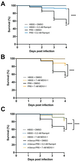
Ramipril and MDIVI-1 increase survival in systemically infected PR8 and Color-flu-infected 3 dpf zebrafish. (A) Increased survival in PR8-infected AB zebrafish treated with 0.2 nM ramipril compared to DMSO controls (**** p < 0.0001). (B) Increased survival in PR8-infected AB zebrafish treated with 7 nM MDIVI-1 compared to DMSO controls (p < 0.0001). (C) Increased survival in mVenus-PR8-infected AB zebrafish treated with 0.2 nM ramipril or 7 nM MDIVI-1 compared to DMSO controls (p < 0.0001). Survival studies were conducted with n = 4 and 50 larvae per sample group.

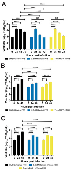
Ramipril and MDIVI-1 treatments lower viral burden in IAV-infected zebrafish. (A) Viral titers for both ramipril (0.2 nM)- and MDIVI-1 (7 nM)-treated 2 dpf zebrafish infected with PR8 were significantly higher at 24 and 48 hpi when compared to 0 hpi, but not different at 72 hpi. For the DMSO-treated zebrafish, viral titers were significantly increased at 24, 48, and 72 hpi (p < 0.0001 for each comparison). For the ramipril-treated zebrafish, viral titers were significantly increased at 24 (p < 0.0001) and 48 hpi (p = 0.0022). For the MDIVI-1-treated zebrafish, viral titers were also significantly increased at 24 and 48 hpi (p < 0.0001 for each comparison). (B) Viral titers for ramipril- and MDIVI-1-treated 3 dpf zebrafish infected with PR8 were significantly lower by 48 hpi (p < 0.0001 for each comparison). (C) Viral titers for ramipril- and MDIVI-1-treated 3 dpf zebrafish infected with mVenus-PR8 were significantly lower at 48 hpi (p < 0.0001 for each comparison). TCID50 assays were conducted using n = 3 and 25 larvae per group for each time point. Not significant (ns), p > 0.05; ** p < 0.01; **** p < 0.0001.

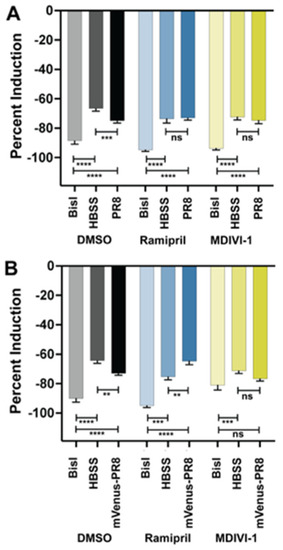
Ramipril and MDIVI-1 treatment alters the respiratory burst response in IAV systemically infected zebrafish at 48 hpi. (A) Respiratory burst response in larvae treated with DMSO, ramipril (0.2 nM), and MDIVI-1 (7 nM) at 48 hpi following systemic injection at 3 dpf with PR8 or HBSS. PR8 infection decreased the response over HBSS in DMSO-treated controls (adjusted p = 0.0008). Both ramipril and MDIVI-1 treatment remedied the reduction in respiratory burst response, as the PR8-infected larvae had the same response as HBSS-injected controls (comparisons were not significant (ns)). The protein kinase C inhibitor bisindolylmaleimide I (BisI) was used as a positive control as it suppresses the respiratory burst response (adj. p < 0.0001 for all comparisons). (B) Respiratory burst response in larvae treated with DMSO, ramipril (0.2 nM), and MDIVI-1 (7 nM) at 48 hpi following systemic injection at 3 dpf with mVenus-PR8 or HBSS. Similar to the PR8-infected larvae, mVenus-PR8 infection decreased the response over HBSS in DMSO-treated controls (adj. p = 0.0040). Ramipril treatment resulted in a higher respiratory burst response with mVenus-PR8 infection than HBSS controls (adj. p = 0.0049). MDIVI-1 treatment remedied the reduction in respiratory burst response as the mVenus-PR8-infected larvae had the same response as HBSS injected controls. The BisI controls were different to the DMSO (adj. p < 0.0001 for both), ramipril (adj. p = 0.0003 for HBSS, and adj. p < 0.0001 for mVenus-PR8), and MDIVI-1 (adj. p = 0.0006 for HBSS, ns for mVenus-PR8) groups. Not significant (ns), p > 0.05; ** p < 0.01; *** p < 0.001; **** p < 0.0001.

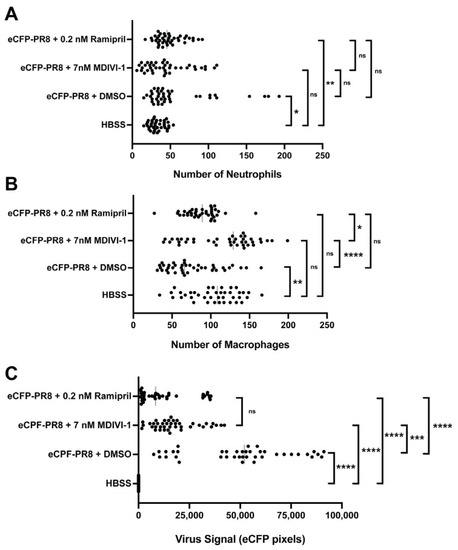
Quantification of the number of neutrophils (A), macrophages (B), and the relative abundance of virus infected cells (C) via fluorescent confocal imaging of Tg(mpeg1:eGFP;lyz:dsRed) larvae at 48 h post injection by eCFP-PR8 or HBSS following treatment with DMSO, ramipril, and MDIVI-1 per optical cross section. For each larva, 10 five-micron optical cross sections were analyzed, which together totaled 50 microns (n = 4 representative larvae). (A) The number of neutrophils increased with eCFP-PR8 infection following DMSO treatment over HBSS controls (adjusted p-value = 0.0145), and with ramipril treatment (adj. p-value = 0.0010), but not with MDIVI-1 treatment. (B) The number of macrophages decreased in eCFP-PR8-infected larvae treated with DMSO over HBSS controls (adj. p-value = 0.0020), but was not different with ramipril or MDIVI-1 treatment. MDIVI-1 treatment increased the number of macrophages compared to the DMSO controls (adj. p-value < 0.0001), and ramipril-treated larvae (adj. p-value = 0.0126). (C) The extent of viral infection was higher in eCFP-PR8-infected larvae treated with DMSO, ramipril, and MDIVI-1 (adj. p-value < 0.0001 for all comparisons). The level of virus infection was lower with ramipril (adj. p-value < 0.0001) and MDIVI-1 (adj. p-value = 0.0008) treatment compared to the DMSO-treated controls. Not significant (ns), p > 0.05; * p < 0.05; ** p < 0.01; *** p < 0.001; **** p < 0.0001.

Acknowledgments
This image is the copyrighted work of the attributed author or publisher, and ZFIN has permission only to display this image to its users. Additional permissions should be obtained from the applicable author or publisher of the image. Full text @ Viruses