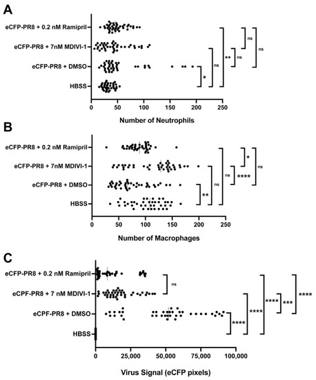
Quantification of the number of neutrophils (A), macrophages (B), and the relative abundance of virus infected cells (C) via fluorescent confocal imaging of Tg(mpeg1:eGFP;lyz:dsRed) larvae at 48 h post injection by eCFP-PR8 or HBSS following treatment with DMSO, ramipril, and MDIVI-1 per optical cross section. For each larva, 10 five-micron optical cross sections were analyzed, which together totaled 50 microns (n = 4 representative larvae). (A) The number of neutrophils increased with eCFP-PR8 infection following DMSO treatment over HBSS controls (adjusted p-value = 0.0145), and with ramipril treatment (adj. p-value = 0.0010), but not with MDIVI-1 treatment. (B) The number of macrophages decreased in eCFP-PR8-infected larvae treated with DMSO over HBSS controls (adj. p-value = 0.0020), but was not different with ramipril or MDIVI-1 treatment. MDIVI-1 treatment increased the number of macrophages compared to the DMSO controls (adj. p-value < 0.0001), and ramipril-treated larvae (adj. p-value = 0.0126). (C) The extent of viral infection was higher in eCFP-PR8-infected larvae treated with DMSO, ramipril, and MDIVI-1 (adj. p-value < 0.0001 for all comparisons). The level of virus infection was lower with ramipril (adj. p-value < 0.0001) and MDIVI-1 (adj. p-value = 0.0008) treatment compared to the DMSO-treated controls. Not significant (ns), p > 0.05; * p < 0.05; ** p < 0.01; *** p < 0.001; **** p < 0.0001.
|
