IMAGE

Figure 3

ID
ZDB-IMAGE-240129-70
Source
Figures for Soos et al., 2024
Image
Figure Caption

Figure 3

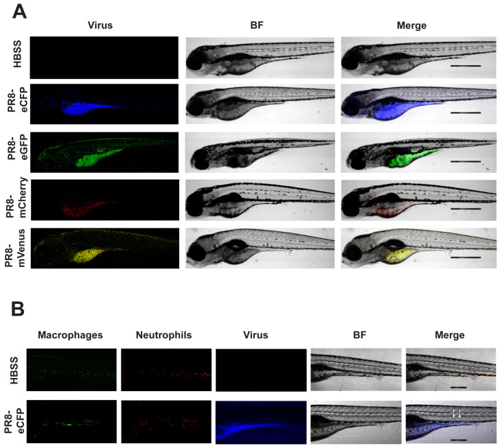
Confocal imaging of Color-flu-infected zebrafish. (A) Representative images of larvae at 24 h post systemic injection of HBSS, eCFP-PR8, eGFP-PR8, mCherry-PR8, and mVenus-PR8 at 3 dpf, at 4× resolution. Scale bar: 300 mm. BF: brightfield. (B) Representative images of Tg(mpeg1:eGFP;lyz:dsRed) larvae at 24 h post systemic injection of HBSS, eCFP-PR8 at 3 dpf at 10× resolution, showing macrophages (green), neutrophils (red), and eCFP-PR8 (blue). Infected skeletal muscle was detected (white arrowheads). Scale bar: 700 μm.

Acknowledgments
This image is the copyrighted work of the attributed author or publisher, and ZFIN has permission only to display this image to its users. Additional permissions should be obtained from the applicable author or publisher of the image. Full text @ Viruses