
- Title
-
Pax4-Ghrelin mediates the conversion of pancreatic ɛ-cells to β-cells after extreme β-cell loss in zebrafish
- Authors
- Yu, J., Ma, J., Li, Y., Zhou, Y., Luo, L., Yang, Y.
- Source
- Full text @ Development
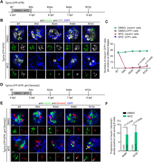
A small proportion of ghrelin-positive ?-cells express insulin after near-total ?-cell ablation. (A) Experimental scheme for DMSO or MTZ treatment of the Tg(ins:CFP-NTR) larvae. (B) Antibody staining of insulin and CFP showed that pancreatic ?-cells in zebrafish regenerated slowly after near-total ablation. (C) The numbers of insulin+ and CFP+ cells per embryo at different stages (n=22). (D) Experimental scheme for DMSO or MTZ treatment of the Tg(ins:CFP-NTR; ghrl:Dendra2) larvae. (E) A small proportion of ?-cells expressed insulin during ?-cell regeneration. Arrowheads indicate CFP+ Dendra2+ double-positive cells. (F) Statistical diagram of proportions of insulin+ Dendra2+ double-positive cells among ?-cells (n=20, the average percentage is shown in the histogram). All statistical data are expressed as mean�s.e.m. P-values were calculated using an unpaired Student's t-test. Scale bars: 10 �m. BT, before treatment; R0 h, R24 h, R48 h and R72 h indicate regeneration for 0, 24, 48 and 72 h, respectively. |
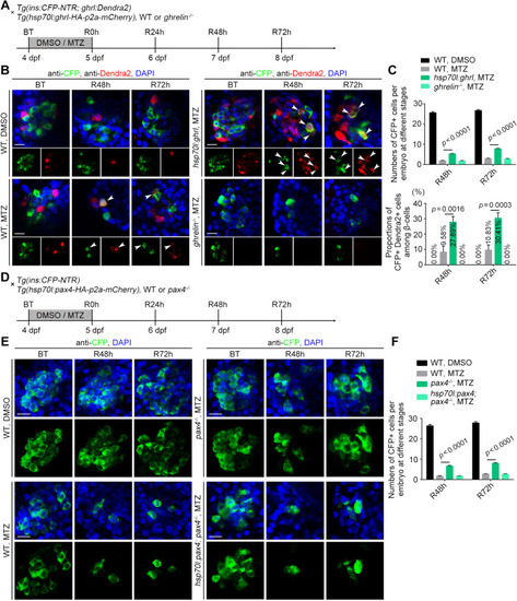
Overexpression of ghrelin or deletion of Pax4 potentiates ?-cell regeneration. (A) Experimental scheme for DMSO or MTZ treatment of the wild-type, ghrelin?/? or Tg(hsp70l:ghrl-HA-p2a-mCherry) larvae under the Tg(ins:CFP-NTR; ghrl:Dendra2) background. (B) Overexpression of ghrelin led to an increased number of neogenic CFP+ cells, including Dendra2+ CFP+ double-positive cells (arrowhead) compared with the wild-type MTZ-treated group, but not in the ghrelin?/? mutant. (C) Numbers of CFP+ cells and proportions of Dendra2+ CFP+ double-positive cells per embryo at different stages (n=20). (D) Experimental scheme for DMSO or MTZ treatment of the wild-type, pax4?/? or Tg(hsp70l:pax4-HA-p2a-mCherry) larvae under the Tg(ins:CFP-NTR) background. (E) The numbers of neogenic CFP+ cells were significantly increased in the pax4?/? mutant, and overexpression of pax4 could rescue the increased ?-cell regeneration in the pax4?/? mutant. (F) The numbers of CFP+ cells per embryo at different stages (n=20). All statistical data are expressed as mean�s.e.m. P-values were calculated using an unpaired Student's t-test. Scale bars: 10 �m. |
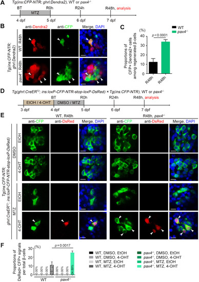
Deletion of Pax4 causes the increased proportion of ?-cells converting to ?-cells during ?-cell regeneration. (A) Experimental scheme for MTZ treatment of the wild-type or pax4?/? mutant under the Tg(ins:CFP-NTR; ghrl:Dendra2) double transgenic background. (B) More neogenic CFP+ ?-cells overlapped with the Dendra2+ ?-cells in pax4?/? mutant relative to the wild-type MTZ-treated group. Arrowheads indicate CFP+ Dendra2+ double-positive cells. (C) Statistical diagram of proportions of CFP+ Dendra2+ double-positive cells among regenerated ?-cells (n=25). (D) Experimental scheme for DMSO, MTZ, ethanol or 4-OHT treatment of the wild-type or pax4?/? mutant under Tg(ghrl:CreERT2; ins:loxP-CFP-NTR-stop-loxP-DsRed; ins:CFP-NTR) triple transgenic background. (E) Antibody staining of CFP and DsRed after 48 h of regeneration (R48 h) showed an increased proportion of DsRed-labelled cells (arrowheads) in the pax4?/? MTZ- and 4-OHT-treated group compared with the wild-type group. (F) The proportions of DsRed+ CFP+ signals per total ?-cells (n=22). All statistical data are expressed as mean�s.e.m. P-values were calculated using an unpaired Student's t-test. Scale bars: 10 �m. |
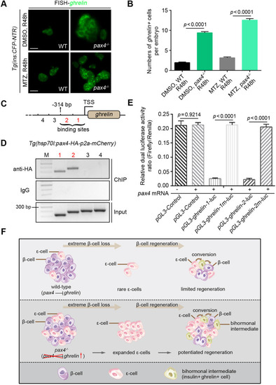
Pax4 binds to genomic DNA at the ghrelin regulatory locus to repress its transcription. (A) Under the Tg(ins:CFP-NTR) transgenic background, fluorescent in situ hybridisation of the ghrelin probe showed that the pax4?/? mutant exhibited a significant expansion of ghrelin-expressing ?-cells both in the DMSO-control and MTZ-treated group compared with the wild-type after 48 h of regeneration (R48 h). (B) Statistical diagram of numbers of ghrelin+ cells per embryo at R48 (n=20). (C,D) Two consecutive sequence fragments 314 bp upstream of the ghrelin TSS were identified as the binding sites of Pax4 (C, fragments 1 and 2), which were shown by ChIP analyses (D, bands 1 and 2). Serial numbers represent the corresponding DNA fragments upstream of the ghrelin TSS. (E) Activities of the luciferase reporter driven by the ghrelin-1 and ghrelin-2 sequences, but not the corresponding mutated sequences (ghrelin-1m and ghrelin-2m), were reduced by pax4 mRNA (n=6). (F) Schematic illustration of the conversion of pancreatic ?-cells to ?-cells mediated by Pax4-ghrelin after extreme ?-cell loss in zebrafish. All statistical data are expressed as mean�s.e.m. P-values were calculated using an unpaired Student's t-test. Scale bars: 10 �m. |