
Fig. 4.
- ID
- ZDB-IMAGE-230420-142
- Genes
- Publication
- Yu et al., 2023 - Pax4-Ghrelin mediates the conversion of pancreatic ɛ-cells to β-cells after extreme β-cell loss in zebrafish
- All Figures
- Figures for Yu et al., 2023
|
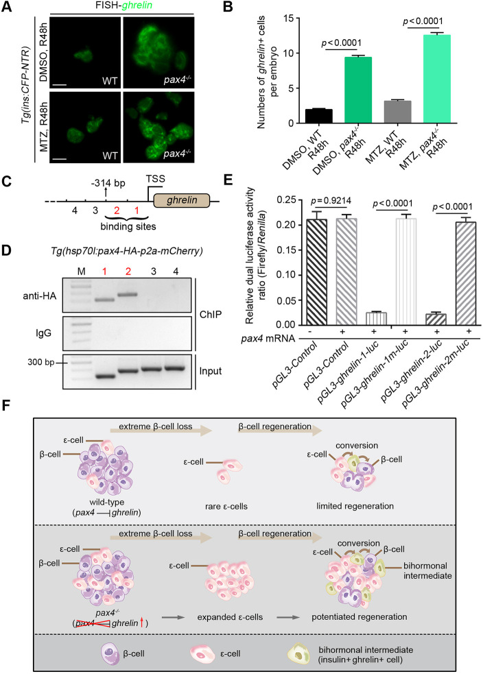
Fig. 4.
Pax4 binds to genomic DNA at the ghrelin regulatory locus to repress its transcription. (A) Under the Tg(ins:CFP-NTR) transgenic background, fluorescent in situ hybridisation of the ghrelin probe showed that the pax4−/− mutant exhibited a significant expansion of ghrelin-expressing ε-cells both in the DMSO-control and MTZ-treated group compared with the wild-type after 48 h of regeneration (R48 h). (B) Statistical diagram of numbers of ghrelin+ cells per embryo at R48 (n=20). (C,D) Two consecutive sequence fragments 314 bp upstream of the ghrelin TSS were identified as the binding sites of Pax4 (C, fragments 1 and 2), which were shown by ChIP analyses (D, bands 1 and 2). Serial numbers represent the corresponding DNA fragments upstream of the ghrelin TSS. (E) Activities of the luciferase reporter driven by the ghrelin-1 and ghrelin-2 sequences, but not the corresponding mutated sequences (ghrelin-1m and ghrelin-2m), were reduced by pax4 mRNA (n=6). (F) Schematic illustration of the conversion of pancreatic ε-cells to β-cells mediated by Pax4-ghrelin after extreme β-cell loss in zebrafish. All statistical data are expressed as mean±s.e.m. P-values were calculated using an unpaired Student's t-test. Scale bars: 10 µm.