
- Title
-
Identification of POLR3B biallelic mutations -associated hypomyelinating leukodystrophy-8 in two siblings
- Authors
- Yang, F., Sun, H., Yang, Y., Wang, Y., Dai, S., Lin, Z., Shen, Y., Liu, H.
- Source
- Full text @ Clin. Genet.
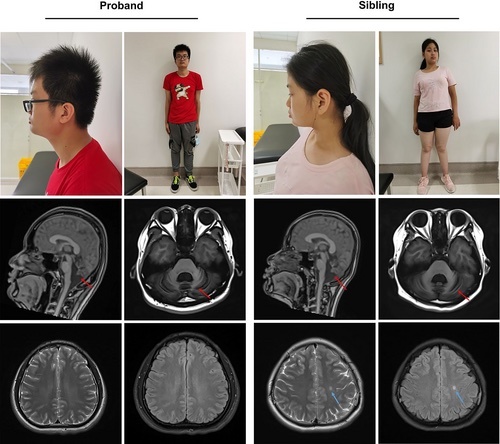
The two patients and brain MRI characteristics. The two patients' characters (row 1). Sagittal and axial T1-weighted images of the brain for two patients at the level of the midline demonstrated vermian cerebellar atrophy (red arrow) (row 2). Axial T2 and FLAIR-weighted images of the brains for two patients showed no significant white matter lesion (row 3). The sibling's MRI showed a sign of potential partial hypomyelination (blue arrow) [Colour figure can be viewed at wileyonlinelibrary.com] |
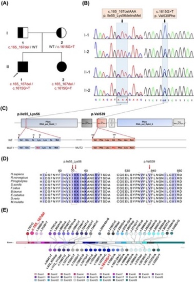
POLR3B biallelic mutations detection in the HLD8 family. (A) The HLD8 family line's genealogy. The proband is represented by the black arrow. (B) Sanger sequence of the variants in POLR3B (c.165_167del; c.1615G>T). The positions of the variants are indicated by black arrows. (C) POLR3B protein domains, localizations of variants, and the comparison of WT amino acids with mutant amino acids. The red lines indicate the positions of the mutations in our study. (D) Conservation of mutant amino acids in different species. (E) The POLR3B pathogenic variants we find (marked in red) in this study differ from pathogenic variants known in POLR3B (Details are available online: https://databases.lovd.nl/shared/transcripts.) [Colour figure can be viewed at wileyonlinelibrary.com] |
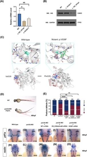
The negative impact of the biallelic variations in POLR3B. (A) The expression of POLR3B mutants by quantitative real-time PCR. Three separate experiments were performed (nonparametric test; *p < 0.05; error bars, s.e.m.). (B) The Western blot analysis of POLR3B expression in cells transfected with plasmids carrying variants. Three independent experiments were performed. (C) PyMol visualization was performed to compare the native and missense mutant amino acid. WT and mutant residues are in light green. (D) Larvae of WT maintained a normal posture. But the polr3b morphants exhibit greater bending angles and body curvature (chi-square test, ****p < 0.0001). (E) Statistics of the embryos with posture-related morphological defects. (F–J) Disruption of polr3b impairs the development of precursor cerebellar Purkinje cells. Typical images of in situ hybridization labeling Purkinje precursors cells with anti-sense probes against ptf1a. (K–O) Disruption of polr3b reduces cerebellar granule cells. Typical images of in situ hybridization labeling cerebellar granule cells with anti-sense probes against reelin [Colour figure can be viewed at wileyonlinelibrary.com]
|