
Fig. 2
- ID
- ZDB-FIG-230830-16
- Publication
- Yang et al., 2023 - Identification of POLR3B biallelic mutations -associated hypomyelinating leukodystrophy-8 in two siblings
- Other Figures
- All Figure Page
- Back to All Figure Page
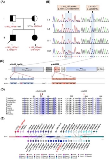
POLR3B biallelic mutations detection in the HLD8 family. (A) The HLD8 family line's genealogy. The proband is represented by the black arrow. (B) Sanger sequence of the variants in POLR3B (c.165_167del; c.1615G>T). The positions of the variants are indicated by black arrows. (C) POLR3B protein domains, localizations of variants, and the comparison of WT amino acids with mutant amino acids. The red lines indicate the positions of the mutations in our study. (D) Conservation of mutant amino acids in different species. (E) The POLR3B pathogenic variants we find (marked in red) in this study differ from pathogenic variants known in POLR3B (Details are available online: https://databases.lovd.nl/shared/transcripts.) [Colour figure can be viewed at wileyonlinelibrary.com] |