
FIGURE
Fig. 1
- ID
- ZDB-FIG-230830-15
- Publication
- Yang et al., 2023 - Identification of POLR3B biallelic mutations -associated hypomyelinating leukodystrophy-8 in two siblings
- Other Figures
- All Figure Page
- Back to All Figure Page
Fig. 1
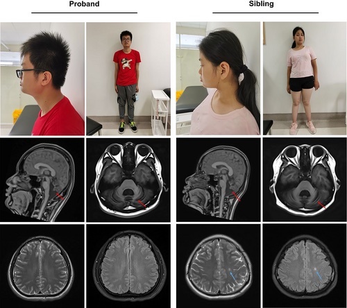
The two patients and brain MRI characteristics. The two patients' characters (row 1). Sagittal and axial T1-weighted images of the brain for two patients at the level of the midline demonstrated vermian cerebellar atrophy (red arrow) (row 2). Axial T2 and FLAIR-weighted images of the brains for two patients showed no significant white matter lesion (row 3). The sibling's MRI showed a sign of potential partial hypomyelination (blue arrow) [Colour figure can be viewed at wileyonlinelibrary.com] |
Expression Data
Expression Detail
Antibody Labeling
Phenotype Data
Phenotype Detail
Acknowledgments
This image is the copyrighted work of the attributed author or publisher, and
ZFIN has permission only to display this image to its users.
Additional permissions should be obtained from the applicable author or publisher of the image.
Full text @ Clin. Genet.