
- Title
-
Changes in Mitochondrial Size and Morphology in the RPE and Photoreceptors of the Developing and Ageing Zebrafish
- Authors
- Burgoyne, T., Toms, M., Way, C., Tracey-White, D., Futter, C.E., Moosajee, M.
- Source
- Full text @ Cells
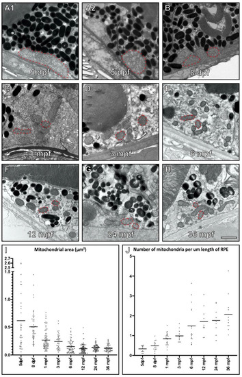
Mitochondria increase in number and decrease in size within the retinal pigment epithelium (RPE) of ageing zebrafish retina. (A–H) Electron microscopy images of mitochondria within zebrafish RPE at 5 dpf through to 36 mpf. The red dotted lines indicate examples of mitochondria within the RPE at each timepoint. All mitochondria were measured in random areas of RPE. When individual mitochondria are examined within the RPE, they (I) decrease in size and (J) increase in number as the zebrafish age. (I,J) At each timepoint, n = 3 zebrafish were examined, and measurements taken from (I) ≥ 34 mitochondria per age group and (J) ≥ 8 regions of the RPE. Statistical significance determined by one way-ANOVA (I–J) with p < 0.0001. Scale (A–G) 1 μm. |
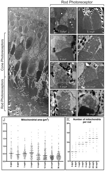
Rod inner segments of early embryonic zebrafish have compact mitochondria with a morphology different from other ages. (A) Electron microscopy image of 12 mpf retina between the RPE and ONL that includes rod and cone photoreceptor outer (POS) and inner segments. (B–I) Images of rod photoreceptor inner segments at 5 dpf through to 36 mpf. (B) At 5 dpf, the mitochondria are bundled together in a spherical like arrangement and there is a morphology change by (C) 8 dpf, with further changes by (D) 1 mpf as the mitochondria become less clumped together. There is (J) a reduction in rod inner segment mitochondrial size and (K) an increase in mitochondria number with time. Measurements were acquired from n = 3 zebrafish and included (J) >83 mitochondria and (K) n = 15 rods at each timepoint. (J,K) Statistical significance determined by one way-ANOVA (J,K) with p < 0.0001. Scale (A) 10 μm, (B–I) 1 μm. |
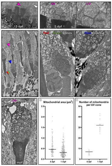
At 5 dpf, the mitochondria within cone inner segments have a morphology that is distinct from those found within 1 month old zebrafish. (A) At 5 dpf, the retina is still developing and it is difficult to distinguish between cone types. UV cones (purple arrowhead) were identifiable due to being positioned furthest from the RPE cell layer. (B) Higher magnification images of UV cone containing packed mitochondria. (C) By 1 mpf, the retina has fully formed and the different photoreceptors can be readily identified. The arrows highlight the inner segment from different photoreceptors: red and green arrows for red and green cones, blue arrows for blue cones, purple arrows for the UV cones and white arrowz for rod photoreceptors. (D–F) The corresponding cone types at 1 mpf are shown at higher magnification. (G) Mitochondria were found to reduce in size and (H) increase in number from 5 dpf to 1 mpf. Measurements were taken from n = 3 zebrafish and included (G) >93 mitochondria and (H) n = 15 UV cones. Statistical significance was determined as (G) p < 0.001 by Mann–Whitney test (non-parametric based on Kolmogorov–Smirnov test) and (H) p < 0.001 by unpaired t-test. Scale (A,C) 5 μm, (B,D–F) 1 μm. |
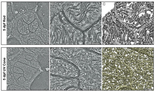
Tomograms show the compact association of mitochondria within the inner segments of UV cone and rod photoreceptors at 5 dpf. (A–C) Tomography data of rod and (D,E) UV cone inner segment mitochondria. (A,D) Single slices from the tomograms with (B,E) higher magnification views. (C–F) Surface rendering of the tomography data allows the architecture of the mitochondria to be assessed in 3D. The rod has elongated and tightly associated cristae membranes, whereas the UV cone has cristae that are wider. Scale bars = 250 nm. |
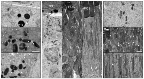
Age-related changes in mitochondrial size in zebrafish are not replicated in developing mouse retinas. (A–C) There is little difference between the mitochondria within the RPE at (A) embryonic day 14 (E14) compared to a (B) postnatal day 13 (P13) and (C) 6 month old mouse (D–F) By E14, mouse retina is still developing and photoreceptors do not have fully formed outer segments (POS). At P13, the mouse has photoreceptors with almost fully developed POS. (G–I) There is a clear difference in the photoreceptor inner segment mitochondria. (G) The mitochondria are smaller at E14 compared to (H) P13 and (I) 6 month old mouse. At P13, the appearance of the (B) RPE and (E, H) photoreceptors are similar to (C,F,I) at 6 months in adult mice. Scale bars (A–I) 1 μm. |
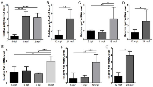
Expression levels of polg2, opa1 and fis1 with age in the zebrafish retina. RT-qPCR was performed to assess expression of polg2 (A,B), opa1 (C,D) and fis1 (E–G) at 5 days post-fertilisation (dpf), 1 month post-fertilisation (mpf), 12 mpf and 24 mpf (n = 5). Expression of fis1 was also assessed at 5–8 dpf (n = 4) (E). * p < 0.05; ** p < 0.01; *** p < 0.001; **** p < 0.0001; n.s, not significant. |
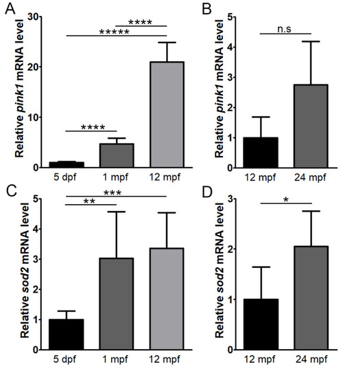
Expression levels of pink1 and sod2 with age in the zebrafish retina. RT-qPCR was performed to assess expression of pink1 (A,B) and sod2 (C,D) at 5 days post-fertilisation (dpf), 1 month post-fertilisation (mpf), 12 mpf and 24 mpf (n = 5). * p < 0.05; ** p < 0.01; *** p < 0.001; **** p < 0.00001; ***** p < 0.0000001; n.s, not significant. |

ZFIN is incorporating published figure images and captions as part of an ongoing project. Figures from some publications have not yet been curated, or are not available for display because of copyright restrictions. |
|
ZFIN is incorporating published figure images and captions as part of an ongoing project. Figures from some publications have not yet been curated, or are not available for display because of copyright restrictions. |