
Figure 3
- ID
- ZDB-FIG-221214-181
- Publication
- Burgoyne et al., 2022 - Changes in Mitochondrial Size and Morphology in the RPE and Photoreceptors of the Developing and Ageing Zebrafish
- Other Figures
- All Figure Page
- Back to All Figure Page
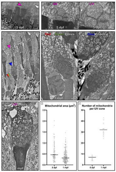
At 5 dpf, the mitochondria within cone inner segments have a morphology that is distinct from those found within 1 month old zebrafish. (A) At 5 dpf, the retina is still developing and it is difficult to distinguish between cone types. UV cones (purple arrowhead) were identifiable due to being positioned furthest from the RPE cell layer. (B) Higher magnification images of UV cone containing packed mitochondria. (C) By 1 mpf, the retina has fully formed and the different photoreceptors can be readily identified. The arrows highlight the inner segment from different photoreceptors: red and green arrows for red and green cones, blue arrows for blue cones, purple arrows for the UV cones and white arrowz for rod photoreceptors. (D–F) The corresponding cone types at 1 mpf are shown at higher magnification. (G) Mitochondria were found to reduce in size and (H) increase in number from 5 dpf to 1 mpf. Measurements were taken from n = 3 zebrafish and included (G) >93 mitochondria and (H) n = 15 UV cones. Statistical significance was determined as (G) p < 0.001 by Mann–Whitney test (non-parametric based on Kolmogorov–Smirnov test) and (H) p < 0.001 by unpaired t-test. Scale (A,C) 5 μm, (B,D–F) 1 μm. |