
- Title
-
Hypoxic and nitrosative stress conditions modulate expression of myoglobin genes in a carcinogenic hepatobiliary trematode, Clonorchis sinensis
- Authors
- Kim, S.H., Yang, D., Bae, Y.A.
- Source
- Full text @ PLoS Negl. Trop. Dis.
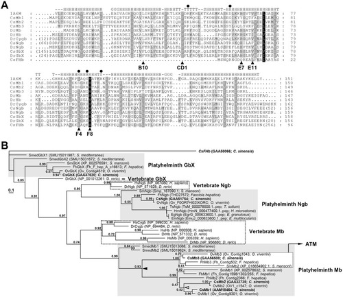
Structural features of the Clonorchis sinensis globins.
A. The amino acid sequences of the C. sinensis proteins were aligned with those of Physeter catodon, of which the tertiary structure was empirically determined (PDB no. 1A6M), and Danio rerio orthologs. The degree of amino acid conservations was highlighted by different shades of grey, and gaps were introduced in the alignment to increase the similarity values. The amino acid positions involved in the binding of heme and oxygen molecules are marked with solid arrowheads and circles (see text). Secondary structure elements determined in the P. catodon globin are shown on the top of the alignment (-, no secondary structure assigned; S, bend; T, turn; G, 3/10-helix; H, alpha-helix). B. A phylogenetic tree of C. sinensis globins and their orthologs. The maximum likelihood tree was constructed using PhyML based on the sequence alignment of globin domains and was rooted in CsFbH. C. sinensis proteins are highlighted in boldface. Branch support values > 0.50, which were obtained by the Shimodaira-Hasegawa-like approximate likelihood ratio test, are indicated at the corresponding branching nodes. The solid arrowhead indicates an evolutionary point where the progenitor of the platyhelminth Mb gene might have been duplicated. |
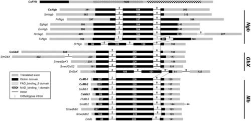
The exon-intron organizations of the Clonorchis sinensis globin genes.
Genomic organizations of the C. sinensis genes and their orthologs were determined by comparing the genomic and transcribed sequences of the respective genes. The coding DNA sequences (CDSs) are represented with gray squares in proportion to their relative sizes and the intervening introns are indicated by solid lines with a fixed length. The phase of each intron, as well as the length of exons and introns in base pairs, is indicated in parentheses. Introns occupying orthologous positions among these genes are connected by vertical dotted lines. The positions of codons encoding functional domains, which are listed in the legend, are differentially marked in the respective CDS regions. |
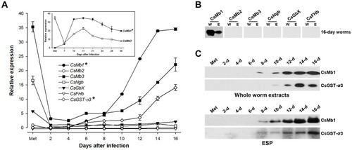
Temporal expression profiles of Clonorchis sinensis globin genes.
A. Induction profiles of the Clonorchis sinensis globin genes were assessed using a quantitative PCR (qPCR) method related to the development/maturation of the liver fluke from metacercariae to 16-day worms. The calculations are based on independent technical triplicates (n = 3, mean ± S.D.). In cases of the CsMb1 and CsMb3 genes, worms up to 56 days-old were included in the measurements (inset graph). The relative expressions of CsMb1 and CsGST-σ3 (marked by asterisks) are given in 1/1,000 of their original values in the graphs. B. Western blot analysis of the whole worm extracts (W, 30 μg/well) of C. sinensis adults and their excretory-secretory products [ESPs (E), 10 μg/well] with antibodies specific to C. sinensis globins. C. The temporal secretion profile of CsMb1 was similarly examined in the whole body extracts and the ESPs of C. sinensis worms from metacercaria to 16 days post-infection. The anti-CsGST-σ3 antibody was also included in the blotting analyses. |
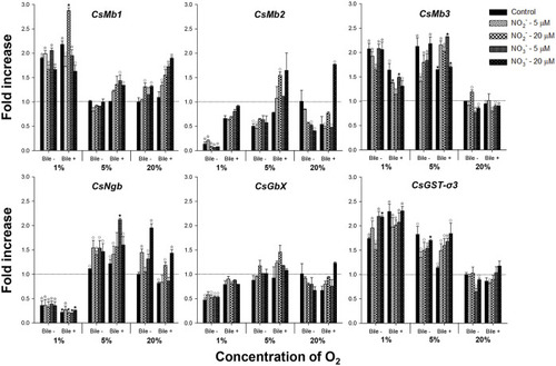
Expression profiles of globin genes in Clonorchis sinensis adults incubated under various experimental conditions.
Live C. sinensis worms were incubated in the RPMI-1640 medium supplemented with bile, nitrite, or nitrate under 1%, 5%, or 20% oxygen conditions. After 24 h incubations, mRNA extracted from the worms was used in qRT-PCR with primer sets specific to each of the C. sinensis globin genes. Fold increases in the globin gene expression in each of the experimental groups were calculated against the 20% oxygen group without any chemical supplement (n = 2, mean ± SD). ○P < 0.05; ☆P < 0.01; ★P < 0.001. |
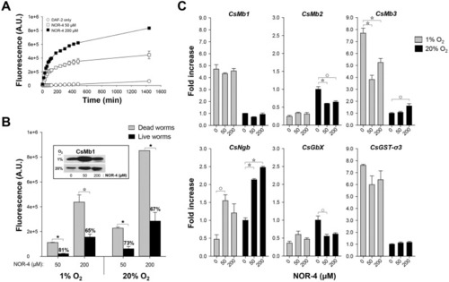
Effect of nitric oxide (NO) on the expressions of the Clonorchis sinensis globin genes.
A. The relative amounts of NO released from NOR-4 in the RPMI-1640 medium was measured using the DAF-2 fluorescence assay method in relation to the incubation times. A.U., arbitrary unit. B. Live or dead C. sinensis adults were incubated for 24 h in the RIMI-1640 medium supplemented with NOR-4 and DAF-2 under 1% or 20% oxygen conditions. The fluorescence values of the conditioned media, which were measured using the DAF-2 method, were serially subtracted from those of NOR-4- and DAF-2-only media (n = 3, mean ± SD). The percentages on the histograms of the live-worm groups indicate the degree of fluorescence reduction compared to those in the dead-worm groups. ☆P < 0.01; ★P < 0.001. The results of western blot analysis of the conditioned media (20 μL/well) with an anti-CsMb1 antibody are shown in the inset image. C. The expression levels of the globin genes were examined in the C. sinensis worms described in panel B by performing qRT-PCR. The fold increases were calculated by comparing the values of the experimental groups with those in the 20% worm-only control group (n = 3, mean ± SD). ○P < 0.05; ☆P < 0.01. |
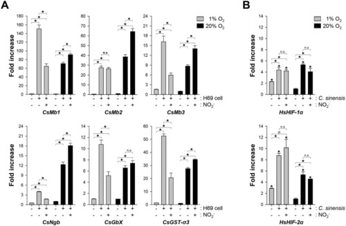
Induction profiles of globin and hypoxia-inducible factor (HIF) genes in co-incubated Clonorchis sinensis adults and H69 cells.
A. The live 28-day-old worms were co-incubated with the human cholangiocytes under the respective conditions. After 24 h incubation, the relative amounts of the C. sinensis gene transcripts were determined using a qRT-PCR method (n = 3, mean ± SD) and the fold increase in each of the experimental groups was calculated against that in the worm-only control group under 20% oxygen condition. B. Fold increase in the expression of HIF-1α and 2α was similarly determined in the human cells. ☆P < 0.01;★P < 0.001; n.s., not significant. |
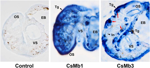
Detection of CsMb1 and CsMb3 in the metacercarial stage of Clonorchis sinensis.
The whole-body sections of C. sinensis metacercariae embedded in paraffin were reacted with mouse antibodies specific to the recombinant forms of C. sinensis CsMb1 and CsMb3. Pooled serum from pre-immune mice (n = 3) was used in the control reaction. The positive reactions were developed into a blue color using a blue chromogen substrate for horseradish peroxidase-conjugated to a second antibody. The red arrows mark a follicle-like structure exhibiting a strong positive signal against the anti-CsMb3 antibody in the subtegumental region. EB, excretory bladder; In, intestine; OS, oral sucker; Tg, tegument; VS, ventral sucker. Original magnifications, x 1,000. |
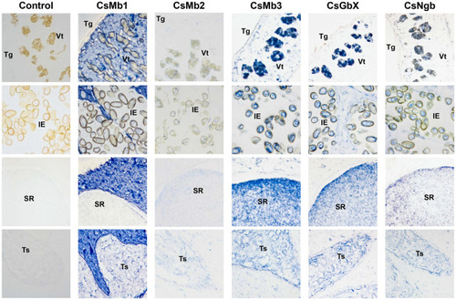
Histological distribution patterns of globins in Clonorchis sinensis adults.
The worm sections were incubated with respective mouse antibodies specific to the C. sinensis globins or a pooled pre-immune mouse serum (n = 3) and then a goat anti-mouse IgG antibody conjugated with horseradish peroxidase (HRP). The specific antigen-antibody reactions were developed into a blue color with a blue chromogen for HRP. IE, intrauterine eggs; SR, seminal receptacle; Tg, tegument; Ts, testis; Vt, vitellaria. Original magnifications, x 200. |
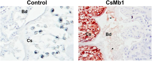
Detection of secreted CsMb1 in the epithelium of rat bile ducts with clonorchiasis.
Histologic sections containing the intrahepatic bile ducts, which were prepared from the liver of an experimental rat with clonorchiasis, sequentially reacted with an anti-CsMb1 antibody or pooled mouse serum (n = 3) and a goat anti-mouse IgG antibody conjugated with horseradish peroxidase (HRP). The positive reactions were visualized by staining with a red chromogen substrate for HRP. The arrowheads indicate examples of the strong positive signal. Bd, epithelium of the bile duct; Cs, Clonorchis sinensis worm body. Original magnifications, x 200. |