
Image
|
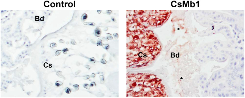
Figure Caption
Fig 9
Detection of secreted CsMb1 in the epithelium of rat bile ducts with clonorchiasis.
Histologic sections containing the intrahepatic bile ducts, which were prepared from the liver of an experimental rat with clonorchiasis, sequentially reacted with an anti-CsMb1 antibody or pooled mouse serum (n = 3) and a goat anti-mouse IgG antibody conjugated with horseradish peroxidase (HRP). The positive reactions were visualized by staining with a red chromogen substrate for HRP. The arrowheads indicate examples of the strong positive signal. Bd, epithelium of the bile duct; Cs, Clonorchis sinensis worm body. Original magnifications, x 200.
Acknowledgments
This image is the copyrighted work of the attributed author or publisher, and
ZFIN has permission only to display this image to its users.
Additional permissions should be obtained from the applicable author or publisher of the image.
Full text @ PLoS Negl. Trop. Dis.