
- Title
-
Sensitivity of Acute Myelocytic Leukemia Cells to the Dienone Compound VLX1570 Is Associated with Inhibition of the Ubiquitin-Proteasome System
- Authors
- Selvaraju, K., Lotfi, K., Gubat, J., Miquel, M., Nilsson, A., Hill, J., Jensen, L.D., Linder, S., D'Arcy, P.
- Source
- Full text @ Biomolecules
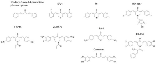
Figure 1. Structures of compounds discussed in the text. |

Figure 2. Time- and dose-dependent loss of viability of MOLM-14 and KG1a cells exposed to VLX1570. Cell viability was determined using acridine orange/propidium iodide staining. |

Figure 3. (A) Accumulation of polyubiquitinated proteins and increased in Hsp70 in KG1a and MOLM14 cells exposed to VLX1570; (B) time- and dose-dependent increased in Grp78/BiP in acute myeloid leukemia (AML) cells exposed to VLX1570; thaps: thapsigargin; (C) VLX1570 induction of proteotoxic stress and ER stress; (D) the loss of cell viability is not affected by the pan-caspase inhibitor z-VAD-fmk (10 μM). Cells were exposed to 0.35 μM VLX1570 for 48 h, and cell viability was determined by acridine orange/propidium iodide staining. The barplots are the average of three replicates of a single experiment. Error bars represent one standard deviation. The experiment was repeated with similar results. |

Figure 5. Depletion of reduced glutathione (GSH) and induction of high-molecular-weight complexes in AML cells. (A) GSH levels in AML cells after different times of exposure to 0.35 μM VLX1570 and/or 10 μM BSO. Means and SDs from one representative experiment are shown; (B) fraction viable cells determined by the AO/PI method after treatment with VLX1570 and/or BSO. Means and SDs from one representative experiment are shown; (C) (top) high molecular weight (HMW) region of Coomassie BB-stained SDS-PAGE gels loaded with extracts from cells exposed for 4 h to different concentrations of VLX1570, (bottom) immunoblots of extracts from VXL1570-exposed cells probed with an antibody to K48-linked polyubiquitin, note the distribution of high molecular weight bands marked with yellow and orange dots; (D) effect of 10 μM BSO on the formation of HMW complexes and loss of HMW proteins in KG1a cells exposed to 0.5, 1, 2, and 5 μM VLX1570; (E) KG1a cell extracts were exposed to 1 μM VLX1570 for increasing times at 37 °C and analyzed by SDS-PAGE; (left): gel stained with Coomassie blue, (right): immunoblot probed with an antibody to K48-linked polyubiquitin; and (F) effect of pretreatment of cell extracts with 20 μM NEM, 100 μM Tiron, or the Michael-acceptor-containing compound CB360 (30 μM) [40] on the formation of HMW complexes. Plots (A,B) show the average of three replicates, and error bars represent one standard deviation. |
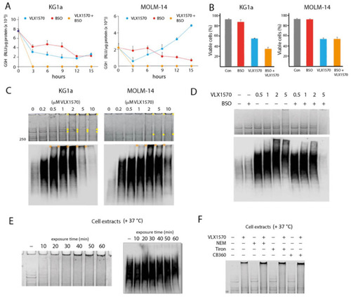
Depletion of reduced glutathione (GSH) and induction of high-molecular-weight complexes in AML cells. ( |
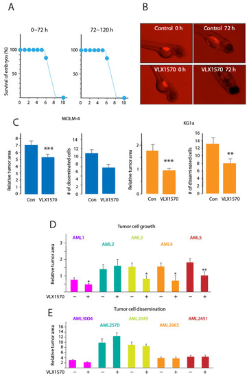
Figure 6. Activity of VLX1570 in the zebrafish embryo model; (A) survival of zebrafish embryos exposed to the indicated concentrations of VLX1570 for 0–72 or 72–120 h; (B) examples of results obtained using labeled AML cells injected into zebrafish embryos followed by treatment with VLX1570; (C) effect of 1 µM VLX1570 on the growth and dissemination of injected AML cell lines in zebrafish embryos (student’s t test, ** p ≤ 0.01, *** p ≤ 0.001); and (D,E) effect of 1 µM VLX1570 on the growth and dissemination of injected primary AML cells in zebrafish embryos (student’s t test, * p ≤ 0.05, ** p ≤ 0.01). |