- Title
-
Lysosomes and the pathogenesis of merosin-deficient congenital muscular dystrophy
- Authors
- Smith, S.J., Fabian, L., Sheikh, A., Noche, R., Cui, X., Moore, S.A., Dowling, J.J.
- Source
- Full text @ Hum. Mol. Genet.
|
Lysosome distribution in muscle biopsies. Cross section of muscle samples from control and MDC1A patients, stained with antibody against Lamp1. Compared with the control sample, lysosomes appear to redistribute within muscle fibers and congregate at the membrane (n = 3 for each). |
|
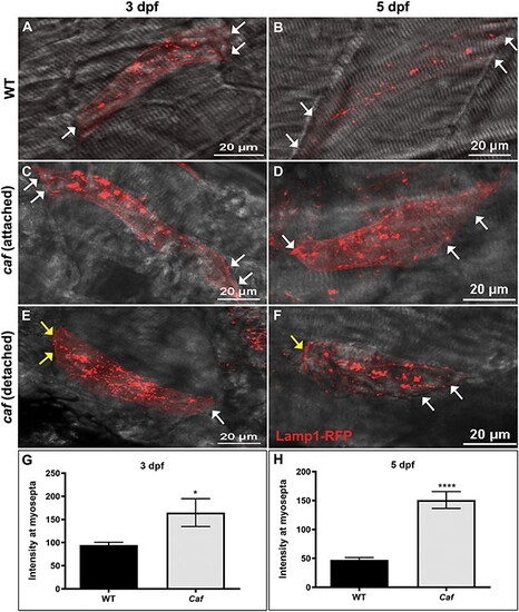
Lysosome distribution in caf mutants. (A–B) Lamp1-RFP expression in WT embryos shows that lysosomes are distributed throughout the cytoplasm in myofibers but not at myosepta (white arrows) at 3 and 5 dpf. (C–F) Lysosomes redistribute partially to the myosepta (white arrows) in caf mutants at 3 dpf (n = 10 fibers) and 5 dpf (n = 25 fibers), in both attached and detached fibers. Yellow arrows indicate where fibers have detached from the myosepta. (G–H) Lamp1-RFP fluorescence intensity at the myosepta was significantly increased in caf mutants at 3 dpf (*, P = 0.035) and 5 dpf (****, P < 0.0001). Bars represent mean ± SEM. PHENOTYPE:
|
|
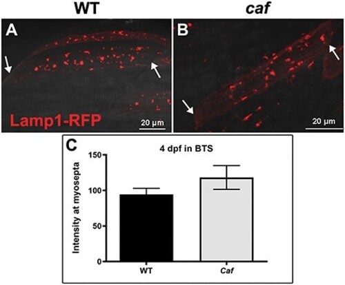
Lysosome distribution in immobilized caf mutants. (A–B) Lysosome distribution in BTS-immobilized WT and caf embryos is similar; lysosomes do not redistribute to the myosepta in paralyzed caf mutants. White arrows indicate the side of the fiber attached to the myosepta. (C) Lamp1-RFP fluorescence intensity at the myosepta was not significantly different in immobilized caf mutants at 4 dpf (P = 0.2591, n = 36 fibers) compared with WT. Bars represent mean ± SEM. |
|
Lysosome distribution in sap mutants. (A) Lamp1-RFP expression in WT embryos shows that lysosomes are distributed throughout the cytoplasm in myofibers but not at myosepta (white arrows) at 4 dpf. (B–C) There is no significant redistribution of lysosomes to the myosepta (white arrows) in sap mutants at 4 dpf (n = 14 fibers), in either attached nor detached fibers. Yellow arrows indicate where fibers have detached from the myosepta. (D) Lamp1-RFP fluorescence intensity at the myosepta was not significantly different in sap mutants at 4 dpf (P = 0.6814, n = 14 fibers) compared with WT. Bars represent mean ± SEM. PHENOTYPE:
|
|
Mean overall Lamp1-RFP fluorescence intensity in muscle fibers. (A) There is an overall increase in Lamp1-RFP expression at 3 dpf (n = 10 fibers) and 5 dpf (n = 25 fibers) in caf mutants compared with WT siblings that is not significant at 3dpf (P = 0.4431) but it is significant at 5 dpf (P = 0.0026). (B) There is no overall difference in Lamp1-RFP expression in immobilized caf mutants compared with immobilized WT siblings (P = 0.3164, n = 36 fibers), nor in sap mutants compared with their WT siblings (P = 0.789, n = 14 fibers). Bars represent mean ± SEM. PHENOTYPE:
|
|
Increasing lysosomal biogenesis moderately protects against myofiber detachment in caf zebrafish. (A) Lysosomal biogenesis transcription factor TFEB increases the number of lysosomes in myofibers, however not significantly (P = 0.1200). (B) Confocal micrographs illustrating GFP-expressing myofibers in WT embryos. (C) Confocal micrographs illustrating GFP-expressing myofibers in caf mutant embryos (detached fibers: TFEB-GFP = 33.9%; PLCδ-PH-GFP = 45.4%; P = 0.0065). |
|
Treatment with the lysosomal agonist MLSA1 partially improves swimming behavior of caf mutants at 5 dpf. (A) MLSA1 reduces swimming behavior of WT zebrafish at 5dpf in a dose-dependent manner. Conversely, in caf mutants, treatment with 5 or 8 μM significantly increases the time spent moving. However, there was no significant improvement in the total distance traveled or average velocity in treated versus untreated caf mutants at any dose tested (n = min 56). Bars represent mean ± SD. (B) Confocal micrographs of whole-mount embryos stained with anti-Lamp1 antibody (green), Phalloidin (magenta) and DAPI (blue). There are no differences in Lamp1 distribution between treated and untreated groups at 5 dpf, and no evidence of increased fiber integrity or decreased detachment. |
|
Autophagy is upregulated in caf mutants but 3-MA treatment does not improve the swimming behavior or dystrophic phenotype of caf mutants. (A) Confocal micrographs of whole-mount embryos stained with anti-LC3 (green), anti-Dystrophin (orange), Phalloidin (magenta) and DAPI (blue). LC3 localization and abundance are affected in caf mutants at 5 dpf; LC3 accumulates at sites of fiber detachment (white arrowheads). (B) Western blot analysis showing increased levels of LC3 II in caf mutants. LC3 II levels were normalized against total protein. (C) Western blot analysis showing decreased levels of LC3 II in WT zebrafish after 3-MA treatment. LC3 II levels were normalized against β-actin. (D–F) Swimming behavior analysis. There was no significant change at 3 dpf in time spent moving (D), total distance traveled (E) or average velocity (F) in caf mutants after treatment with 10 mM 3-MA (n = 165). Bars represent mean ± SEM. PHENOTYPE:
|
|
Vps34-IN1 treatment does not significantly improve the swimming behavior and dystrophic phenotype of caf mutants. (A and B) There is a small magnitude, non-significant, increase in time spent moving and total distance traveled in caf mutants after treatment with 100 nM Vps34-IN1 (n = 96). (C) There is no change in average velocity in caf mutants after treatment with 100 nM Vps34-IN1 (n = 96). (D) Birefringence intensity measurements show no significant improvement to the dystrophic phenotype of caf mutants after Vps34-IN1 treatment (n = 16). ****P < 0.0001. Bars represent mean ± SEM. |
|
UPS is upregulated and MG-132 treatment improves the swimming behavior and dystrophic phenotype of caf mutants. (A and B) Western blot analysis showing increased levels of Polyubiquitinylated proteins (PolyU) in caf mutants. (C–E) Swimming behavior analysis. (C) There is no significant improvement in time spent swimming in caf mutants treated with 10 μM MG132 (n = 166). (D and E) There are significant improvements in distance traveled and average velocity in caf mutants at 3 dpf after treatment with 10 μM MG132. (F) Treatment with 10 μM MG132 improves the dystrophic phenotype of caf mutants at 3 dpf but does not restore muscle integrity to WT levels (n = 43). **P < 0.0075; ****P < 0.0001. Bars represent mean ± SEM. PHENOTYPE:
|


