
Fig. 4
- ID
- ZDB-IMAGE-230317-4
- Publication
- Smith et al., 2021 - Lysosomes and the pathogenesis of merosin-deficient congenital muscular dystrophy
- All Figures
- Figures for Smith et al., 2021
|
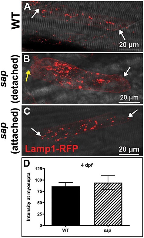
Fig. 4 Lysosome distribution in sap mutants. (A) Lamp1-RFP expression in WT embryos shows that lysosomes are distributed throughout the cytoplasm in myofibers but not at myosepta (white arrows) at 4 dpf. (B–C) There is no significant redistribution of lysosomes to the myosepta (white arrows) in sap mutants at 4 dpf (n = 14 fibers), in either attached nor detached fibers. Yellow arrows indicate where fibers have detached from the myosepta. (D) Lamp1-RFP fluorescence intensity at the myosepta was not significantly different in sap mutants at 4 dpf (P = 0.6814, n = 14 fibers) compared with WT. Bars represent mean ± SEM.