FIGURE SUMMARY
Title

Leucyl-tRNA synthetase deficiency systemically induces excessive autophagy in zebrafish

Authors
Inoue, M., Miyahara, H., Shiraishi, H., Shimizu, N., Tsumori, M., Kiyota, K., Maeda, M., Umeda, R., Ishitani, T., Hanada, R., Ihara, K., Hanada, T.
Source
Full text @ Sci. Rep.

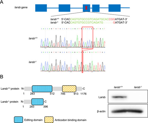
ZFIN is incorporating published figure images and captions as part of an ongoing project. Figures from some publications have not yet been curated, or are not available for display because of copyright restrictions.

Construction of larsb-knockout mutant zebrafish line. (A) Diagram showing the larsb genomic locus, CRISPR/Cas9 target site, and larsb-knockout (larsb−/−) zebrafish mutant genotype. The sgRNA target sequence is displayed in green and the PAM region in red. In the genomic sequencing analysis chromatograms, the deletion region in the mutant larsb−/− zebrafish is shown by the red box. (B) The Lars protein of larsb−/− zebrafish had a missing editing domain. Western blot analysis of the Larsb protein expression in larsb+/+ and larsb−/− zebrafish. β-actin levels served as the loading control. Larsb: leucyl-tRNA synthetase b.

EXPRESSION / LABELING:
Antibody:
Fish:
Anatomical Term:
Stage: Protruding-mouth
PHENOTYPE:
Fish:
Observed In:
Stage: Protruding-mouth

Larsb-knockout larvae display severe developmental phenotype and liver abnormality with early lethality. (A) Bright field lateral views of larsb+/+ and larsb−/− embryos at 96 h post fertilization (hpf). Scale bar: 500 μm (top row) and 300 μm (bottom row). (B) Kaplan–Meier survival curve of larsb+/+ (n = 32), larsb+/- (n = 63), and larsb−/− (n = 23) larvae. (C) Lateral views of larsb+/+ and larsb−/− embryos containing haemoglobin-containing cells (white arrows) stained with o-dianisidine at 72 hpf. (D) Morphological abnormality at 3 dpf and 6 dpf in the livers of larsb−/− larvae under Tg[fabp10:mcherry] background. Scale bar: 300 μm. (E) Quantification of liver size in larsb−/− larvae under Tg[fabp10:mcherry] background (3 dpf and 6 dpf). Liver sizes were evaluated using ImageJ software version 1.52a (https://imagej.nih.gov/ij/). n = 5 fish/group. Error bars indicate SEM. Student’s t-test; ***P < 0.001. Statistics were calculated and the figure was produced in GraphPad software version 8 (https://www.graphpad.com/scientific-software/prism/). Larsb: leucyl-tRNA synthetase b, dpf: days post fertilization.

Histopathology and fluorescent immunostaining of larsb-knockout larvae. (A) Lower magnification sagittal views (top row) and higher magnification views (bottom row) of larsb+/+ and larsb−/− larvae. Huge vacuolations, which seemed to disappear in the cytoplasm, were seen in the livers of larsb−/− larvae (black arrows), and some large vacuolations included a bare nucleus (black arrowheads). Scale bar: 25 µm. (B) Western blot analysis of p62 and Lc3b protein expression in larsb+/+ and larsb−/− larvae. β-actin levels served as the loading control. (C) Lower magnification sagittal views of larsb+/+ and larsb−/− larvae (top row). Fluorescent immunostaining against Lc3b (green) and DAPI (blue) of the livers, skeletal muscles, and spinal cords of larsb+/+ and larsb−/− larvae. Livers of larsb−/− larvae had large vacuoles, including floating nuclei and various sized dots with Lc3b immunoreactivity (white arrows). Skeletal muscles and spinal cords of larsb−/− larvae had many dots with Lc3b immunoreactivity (white arrowheads). Scale bar: 10 µm. Lars: leucyl-tRNA synthetase; Lc3b: microtubule-associated protein 1A/1B-light chain 3; DAPI: 4′,6-diamidino-2-phenylindole.

Immunoelectron microscopy of larsb-knockout larvae under Tg[fabp10:mcherry] background. (A–C) Immunoelectron microscopy of the liver, skeletal muscle, and spinal cord of larsb+/+ larva. (D–I) Immunoelectron microscopy of the liver, skeletal muscle, and spinal cord of larsb−/− larva. The bottom row shows higher magnification images (G–I). Large vacuoles in the livers of larsb−/− larvae (asterisks) were composed of numerous irregular membranous structures, which showed immunoreactivity against both Lc3b (black arrows) and mCherry (black arrowheads) (D,G). Scale bar: 5.0 µm for (A,D); 1.0 µm for B and E; 500 nm for (C,F,H); 100 nm for (G,I). AP: autophagosome, N: nucleus, Larsb: leucyl-tRNA synthetase, Lc3b: microtubule-associated protein 1A/1B-light chain 3.

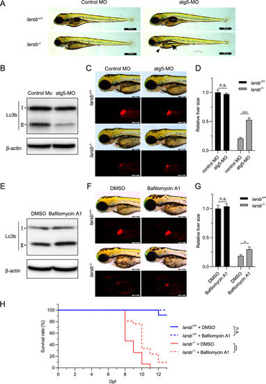
Inhibition of autophagy prevents abnormal development and improves survival in larsb-knockout larvae. (A) Morphology of larsb+/+ and larsb−/− embryos injected with either control MO or atg5-MO (72 h post fertilization (hpf)). Scale bars: 500 µm. (B) Western blot analysis of Lc3b protein expression at 72 hpf for wild-type embryos injected with either control MO or atg5-MO. β-actin levels served as the loading control. (C) Morphological abnormality at 72 hpf in the livers of larsb−/− larvae under Tg[fabp10:mcherry] background injected with either control MO or atg5-MO. Scale bars: 200 μm. (D) Quantification of liver size in larsb−/− larvae under Tg[fabp10:mcherry] background (72 hpf). Liver sizes were evaluated using ImageJ software version 1.52a (https://imagej.nih.gov/ij/). n = 4 fish/group. Error bars indicate SEM. Student’s t-test; ***P < 0.001. (E) Western blot analysis of Lc3b protein expression at 72 hpf for wild-type embryos treated with DMSO or bafilomycin A1. β-actin levels served as the loading control. (F) Morphological abnormality at 72 hpf in the livers of larsb−/− larvae under Tg[fabp10:mcherry] background treated with DMSO or bafilomycin A1. Scale bars: 200 μm. (G) Quantification of liver size in larsb−/− larvae under Tg[fabp10:mcherry] background (72 hpf). Liver sizes were evaluated using ImageJ software version 1.52a (https://imagej.nih.gov/ij/). n = 10 fish/group. Error bars indicate SEM. Student’s t-test; *P < 0.05. (H) Kaplan–Meier survival curve of larsb+/+ (n = 23) and larsb−/− (n = 15) larvae treated with DMSO and larsb+/+ (n = 11) and larsb−/− larvae (n = 21) treated with bafilomycin A1. Statistics were calculated and the figure was produced in GraphPad software version 8 (https://www.graphpad.com/scientific-software/prism/). Larsb: leucyl-tRNA synthetase b, MO: morpholino, n.s.: non-significant, DMSO: dimethyl sulfoxide, Dpf: days post fertilization.

Acknowledgments
This image is the copyrighted work of the attributed author or publisher, and ZFIN has permission only to display this image to its users. Additional permissions should be obtained from the applicable author or publisher of the image. Full text @ Sci. Rep.