
Figure 4
- ID
- ZDB-IMAGE-210424-5
- Antibodies
- Publication
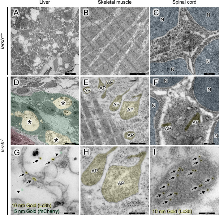
- Inoue et al., 2021 - Leucyl-tRNA synthetase deficiency systemically induces excessive autophagy in zebrafish
- All Figures
- Figures for Inoue et al., 2021
|
Figure 4
Immunoelectron microscopy of