
- Title
-
Chaga mushroom (Inonotus obliquus) polysaccharides exhibit genoprotective effects in UVB-exposed embryonic zebrafish (Danio rerio) through coordinated expression of DNA repair genes
- Authors
- Eid, J.I., Al-Tuwaijri, M.M., Mohanty, S., Das, B.
- Source
- Full text @ Heliyon
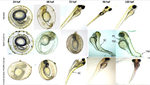
Bright field images of morphological analysis of zebrafish embryos (24 hpf to 120 hpf) in the three different zebrafish groups (control, UVB-exposed, IOP treated UVB-exposed). The structural deformities are shown in arrows: SC-spinal curvature, PE-pericardial edema, YSE-yolksac edema, TM-tail malformation. |
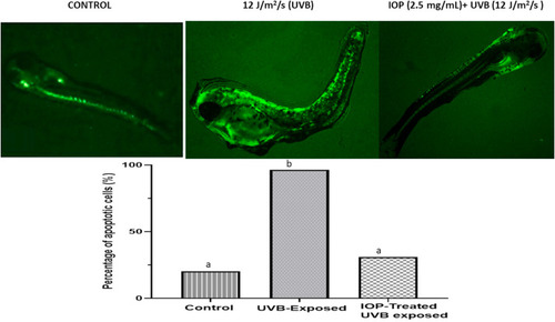
Acridine orange (AO) staining of zebrafish embryos (5 dpf): Above: Acridine orange staining pics for 5dpf zebrafish for the three groups; control, UVB-exposed, IOP treated UVB-exposed, showing intensive green fluorescence as significant uptake of AO dye due to cellular and DNA damage. Below: Histogram generated by Image J analysis showing the percentage of apoptotic cells in the three groups. Apoptosis was significantly high in the UVB-exposed group in comparison to control and IOP-treated UVB-exposed group. Different letters represent statistically significant differences (ANOVA, P < 0.0001). |
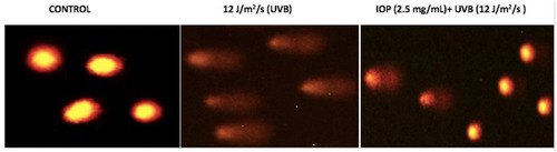
Alkaline comet assay showing distinct comet head and tail after ethidium bromide staining and fluorescent microscopy in the three zebrafish groups control, UVB-exposed, IOP treated UVB-exposed at 5dpf. |
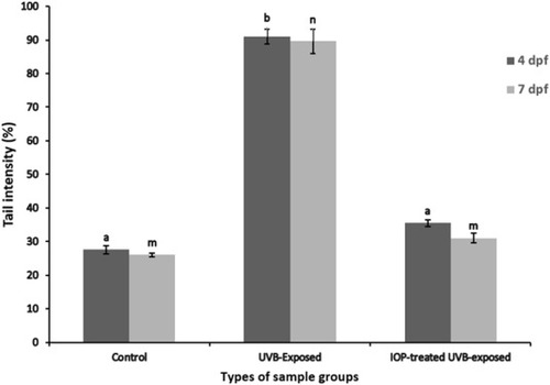
Comet assay histogram at 5 dpf and 7 pdf generated by analyzing the tail intensity (DNA fragmentation) using Image J in the three zebrafish (groups control, UVB-exposed, IOP treated UVB-exposed). Tail intensity was significantly increased in the UVB-exposed group compared to control and IOP-treated UVB exposed groups (ANOVA at 4dpf, F = 2021, p < 0.0001, ANOVA at 7 dpf F = 2256, p < 0.0001). Different letters represent statistically significant differences (P < 0.05). |
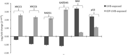
qRT-PCR bar graphs showing the log2 fold change in the gene expression in UVB-exposed and IOP-treated UVB exposed groups (5 dpf) compared to control (set at 0). Significant upregulation was observed in GADD45, RAD51, XRCC5, and XRCC6 in the IOP-treated UVB-exposed group (Kruskal-Wallis test, H = 32.31, p < 0.001) compared to only UVB-exposed group. UVB exposed group showed significant upregulation of p53 and BAX genes (p < 0.0001) that indicated enhanced apoptosis and cell death leading in zebrafish embryos. |
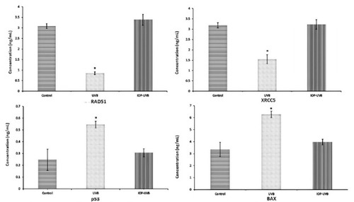
Sandwich ELISA analysis depicting the concentration of XRCC5, RAD51, p53, and BAX in control, UVB-exposed and IOP-UVB-exposed zebrafish larval pools (7 dpf, 10 larvae/pool/group). The levels of XRCC5 and RAD51 were significantly decreased inUVB exposed group compared to IOP-treated UVB-exposed group, whereas p53 and BAX levels were significantly increased in the UVB-exposed groups. Data is shown for a total of 120 zebrafish larvae/group. The readings were recorded in duplicates at 450 nm. ∗ signifies a statistical significant value at p < 0.05. |
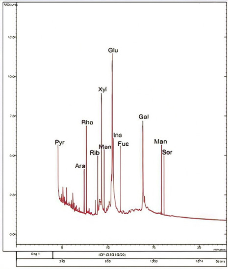
GC-MS chromatogram depicting the retention time peaks for different monomers of Chaga mushroom polysaccharides after hot water ethanolic extraction. |
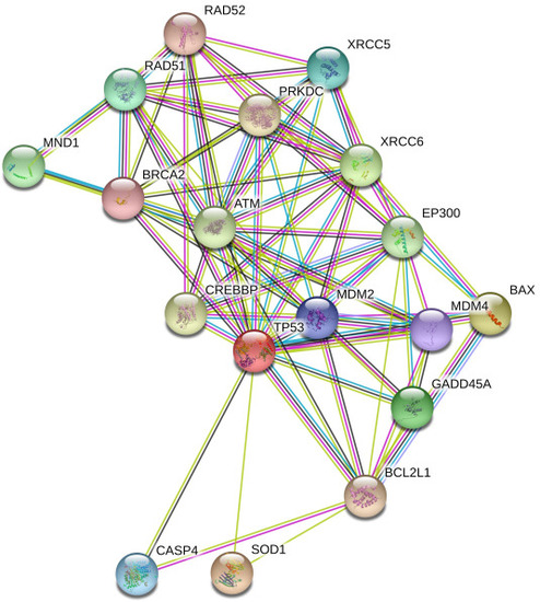
Network analysis using String software demonstrating the coordinated association of the DNA repair genes involved during UV induced DNA repair. DNA repair genes such as RAD51, XRCC5, XRCC6, and GADD45 acted collectively and were involved in coordinate expression. p53 was the central player involved in inducing the expression of pro-apoptotic gene, BAX. |