FIGURE SUMMARY
Title

Wnt-controlled sphingolipids modulate Anthrax Toxin Receptor palmitoylation to regulate oriented mitosis in zebrafish

Authors
Castanon, I., Hannich, J.T., Abrami, L., Huber, F., Dubois, M., Müller, M., van der Goot, F.G., Gonzalez-Gaitan, M.
Source
Full text @ Nat. Commun.

Wnt5b regulates sphingolipid levels in zebrafish embryos.

ad, f Volcano plots of lipidomic data for wnt5b morphants compared to control embryos at gastrula stage (6hpf at 28 °C) from four independent experiments. Horizontal dashed line thresholds significance (p-value = 0.05). NS (statistically non-significant); S (statistically significant). Unpaired, two-tailed student’s t-test was used. Blue shadowed area delimits +/−1.5-fold difference in abundance with respect to control. Relevant changes in the decrease of the content of a lipid species are located in the upper left quadrant. Each dot corresponds to an individual lipid species. a, c Sphingolipids in deyolked wnt5b morphant embryos (a) or in the yolk cell of wnt5b MO-injected embryos (c). Blue dots correspond to ceramides (Cer), red to Hexosylceramides (HexCer) and yellow to sphingomyelin (SM). b, d Phospholipids (PC, PE, PI, and PS) in deyolked wnt5b morphant embryos (b) or in the yolk cell of wnt5b MO-injected embryos (d). Note that the phospholipids significantly downregulated or upregulated beyond 1.5-fold in b and d correspond to phospholipids present at very low levels (between 1.3 x 10−3 and 8.4 x 10−7 Mol%) and therefore their amounts cannot be precisely determined in control versus wnt5b morphant conditions. e Simplified scheme representing both the de novo and salvage pathways for sphingolipid biosynthesis, with selected metabolites and genes indicated. DHCer dihydroceramide, HexDHCer dihydrohexosylceramide, DHSM dihydrosphingomyelin. f Volcano plot for sphingolipids in the de novo pathway in wnt5b deyolked morphant embryos. Lipid amounts are normalized as pmol/nmol inorganic phosphate (see “Methods”). Source data are provided as a Source Data file.

PHENOTYPE:
Fish:
Observed In:
Stage: Shield

Wnt5b signaling regulates the levels of sphingolipids by modulating SPT activity.

a Scheme of metabolic tracer assay. One-cell stage embryos were injected with (13C315N)-l-serine (heavy serine/serine*), which is incorporated into 3-ketosphinganine by SPT, losing one heavy carbon. Incorporation of the remaining three heavy carbons into the downstream sphingoid base sphingosine is used as a direct in vivo readout for SPT activity. Heavy sphingosine (red) co-elutes with sphingosine (black), but it can be distinguished due to its higher mass/charge ratio (m/z). These experiments were done three times independently for each condition. b, c Relative amounts of heavy sphingosine in wnt5a morphant embryos (b), wnt5b morphants (c) or embryos treated with Myriocin (b, c) from three independent experiments. d, e Relative amounts of C18 sphinganine (d) or C18 sphingosine (e) in wnt5a morphant embryos from three independent experiments. f, g Relative amounts of C18 sphinganine (f) or C18 sphingosine (g) in wnt5b morphant embryos from three independent experiments. Unpaired, two-tailed student’s t-test was used in (bg). All error bars represent the standard error of the mean (SEM). hk Orientation of division of epiblast cells with respect to the A/V embryonic axis determined by using H2A.F/Z:GFP transgenic line to monitor chromosome separation during anaphase. Left, representative confocal images of dorsal epiblast cells expressing H2A-GFP in wnt5a morphants (h), in wnt5a morphants treated with sphinganine (0.5 nl of 10 μM) (i), in wnt5b morphants (j) and in wnt5b morphants treated with sphinganine (0.5 nl of 25 μM) (k). Division axes are marked by arrows. Animal pole is up. Scale bars: 10 µm. Right, polar graphs showing the frequency distribution of the angle between the division axis and the A/V embryonic axis in wnt5a morphants (h) and in wnt5a morphants treated with sphinganine (0.5 nl of 10 μM) (i), wnt5b morphants (j) and in wnt5b morphants treated with sphinganine (0.5 nl of 25 μM) (k). n (number of cell divisions analyzed) over N (number of embryos). Note that rescue of randomization of division in wnt5b morphants requires higher amounts of sphinganine than for wnt5a morphants. In (hk), χ2 test was used (see Supplementary Table 1). Source data are provided as a Source Data file.

<italic>sptlc1</italic>-deficient embryos exhibit low levels of sphingolipids.

a Structure of zebrafish sptlc1 gene. Single-guide RNA used is shown in green. The sequence deleted in the CRISPR/Cas9 mutant is shown in red in the annotated sptlc1 sequence. b Graph showing the relative amounts of C18 sphinganine in 4 dpf sptlc1ug105/ug105 normalized to the average of the amounts of C18 sphinganine in their siblings. Data are represented as the mean value +/- SEM from three independent experiments. Unpaired, two-tailed student’s t-test was used. cf Volcano plots of lipidomic data from three (cd) or two (e, f) independent experiments showing either sphingolipids in deyolked embryos for sptlc1 morphants at gastrula stages (c) or in 4 dpf sptlc1ug105/ug105 mutants (e) or phospholipids in deyolked embryos for sptlc1 morphants at gastrula stages (d) and 4 dpf sptlc1ug105/ug105 mutants (f). Horizontal dashed line thresholds significance (p-value = 0.05). NS (statistically non-significant); S (statistically significant). An unpaired, two-tailed student’s t-test was used for analysis of sptlc1 morphants. A paired, two-tailed student’s t-test was used for sptlc1 mutants. Blue shadowed area delimits +/–1.5-fold difference abundance with respect to control. Relevant changes in the decrease of the content of a lipid species are located in the upper left quadrant. Each dot corresponds to an individual lipid species. Note that phospholipids downregulated in deyolked sptlc1 morphants and upregulated in sptlc1ug105/ug105 mutants correspond mainly to PE and minor PI species (for details see Supplementary Fig. 5e, h). Lipid amounts were normalized as pmol/nmol inorganic phosphate (see “Methods”). Source data are provided as a Source Data file.

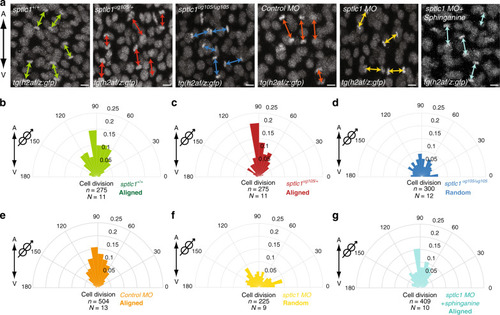
Sphingolipids regulate oriented divisions of epiblast cells.

ag Orientation of division of dorsal epiblast cells with respect to the A/V embryonic axis determined by using H2A.F/Z:GFP transgenic line to monitor chromosome separation during anaphase. a Representative confocal images of dorsal epiblast cells expressing H2A-GFP in sptlc1+/+, sptlc1ug105/+ and sptlc1ug105/ug105 embryos and in control, sptlc1 morphants and sptlc1 morphant embryos treated with sphinganine (from left to right) from at least nine independent experiments. Division axes are marked by arrows. Animal pole is up. Scale bars: 10 µm. bg Polar graphs showing the frequency distribution of the angle between the division axis and the A/V embryonic axis from (a) in sptlc1+/+, (b) sptlc1ug105/+, (c) sptlc1ug105/ug105, (d) control morphants, (e) sptlc1 morphants, (f) and sptlc1 morphants treated with sphinganine (g). n (number of cell divisions analyzed) over N (number of embryos). Divisions are aligned along the A/V axis in sptlc1+/+, sptlc1ug105/+ and in control morphant embryos, but randomized in sptlc1ug105/ug105 embryos and sptlc1 morphants. Randomization of division in sptlc1 morphants is rescued by injection of 0.5 nl of 0.25 μM sphinganine. In (bg), χ2 test was used (see Supplementary Table 1). Cartoons at the corner of polar graphs in this report indicate the parameter measured. Source data are provided as a Source Data file.

Formation of F-actin and Antxr2a caps are sphingolipid-independent.

a Scheme representing the three functions of the actin cytoskeleton during oriented divisions of epiblast cells5. In metaphase, epiblast cells form a polarized F-actin cap aligned with respect to the A/V axis. The formation of this F-actin cap depends on the small GTPase RhoA. The transmembrane protein Antxr2a binds to actin, and it is therefore recruited to and enriched at the actin cap to form a cap itself. Both caps colocalize. Antxr2a interacts then with an active form of RhoA to activate Diaphanous, which connects the mitotic spindle with the caps, rotating the spindle to align it with the caps and therefore with the embryonic axis. As a result, divisions of epiblast cells are oriented along the A/V embryonic axis. b, e Representative confocal images from a time-lapse movie of epiblast cells at metaphase (META) and anaphase (ANA) expressing the F-actin biosensor (Lifeact-mRFP) (b) or Antxr2a-eYFP (e) in control embryos (left panels) or sptlc1 morphants (right panels) from at least ten independent experiments. Yellow dashed line highlights F-actin or Antxr2a cap. Green dashed line, division plane. Animal pole, up. The embryonic axis is represented by a bi-directional arrow on the right. Scale bars: 10 µm. c Quantification of cortical F-actin accumulation at the cap of epiblast cells in control (n = 19 cells; green) and sptlc1 morphants (n = 15 cells; red) expressing Lifeact-mRFP. d, g Polar graphs showing the frequency distribution of the angle between the F-actin cap (d) or the Antxr2a cap (g) and the plane of the A/V embryonic axis in control (left graph) and sptcl1 morphants (right graph). n (number of cells analyzed) over N (number of embryos). In (d, g), χ2 test was used (see Supplementary Table 1). f Antxr2a accumulation at the cap in epiblast cells expressing WT-Antxr2a-eYFP in control (n = 24 cells; green) or in sptlc1 morphants (n = 15 cells; red) and expressing AAC-zAntxr2a-eYFP in control morphants (n = 19 cells; blue). Data in (c, f) are represented as the mean value +/− SEM and an unpaired, two-tailed student’s t-test was used. DP (Division plane). Source data are provided as a Source Data file.

Alignment of F-actin and Antxr2a caps with the division plane requires sphingolipids.

a, c Polar graphs showing the frequency distribution of the angle between the F-actin cap (a) or the Antxr2a cap (c) with respect to the mitotic plane (green line in Fig. 5b, e). b, d Polar graphs showing the frequency distribution of the angle between the plane of division and the plane of the embryonic axis in epiblast cells expressing either Lifeact-mRFP biosensor (b) or Antxr2a-eYFP (d) in control and sptlc1 morphant embryos. χ2 test was used (see Supplementary Table 1). n (number of cells analyzed) over N (number of embryos). DP division plane. Source data are provided as a Source Data file.

Sphingolipids regulate Antxr2a palmitoylation to control oriented divisions.

a, c3H-palmitate incorporation assay. Representative blots showing 3H-palmitate levels incorporated into WT-zAntxr2 or AAC-zAntxr2a expressed in HeLa cells (a) or into WT-zAntxr2a expressed in either Control, SPT, WNT5a or WNT5b RNAi HeLa cells (c). b, d Quantification of 3H-palmitate incorporation as shown in (a, c), normalized to the average of incorporation in WT-zAntxr2a from three independent experiments. Silencing SPT or WNT5b results in a reduction of Antxr2a palmitoylation levels. WNT5a silencing causes a mild, but statistically significant effect on receptor palmitoylation. Data are represented as the mean value +/– SEM. Unpaired, two-tailed student’s t-test was used. eh Division orientation of epiblast cells with respect to the A/V embryonic axis. e, f Representative confocal images of epiblast cells expressing H2A-GFP in sptlc1 morphants (e) or in sptlc1 morphants treated with palmostatin B (f) from at least eight independent experiments. Division axes are marked by arrows. Animal pole is up. Scale bars: 10 µm. g, h Polar graphs showing the frequency distribution of the angle between the division axis and the A/V axis in sptlc1 morphants and sptlc1 morphants treated with palmostatin B. χ2 test was used (see Supplementary Table 1). i Representative confocal images from a time-lapse movie of a dorsal epiblast cell at metaphase (META) and anaphase (ANA) expressing AAC-Antxr2a-eYFP in control morpholino-injected embryos. Yellow dashed line corresponds to AAC-Antxr2a cap. Green dashed line, division plane (DP). Embryos are oriented with the animal pole up. Embryonic axis is represented by a bi-directional arrow on the right. Scale bars: 10 µm. jm Polar graphs showing the frequency distribution of either the angle between the division plane (green dashed line in i) and the A/V embryonic plane (j), the angle between the Antxr2a cap and the A/V embryonic axis (k) or the angle between the cap and the division plane of epiblast cells in live embryos expressing AAC-Antxr2a-eYFP (l) or expressing AAC-Antxr2a and ΔBDG-mDia2 (m). χ2 test was used (see Supplementary Table 1). n (number of cells analyzed) over N (number of embryos). Source data are provided as a Source Data file.

ZFIN is incorporating published figure images and captions as part of an ongoing project. Figures from some publications have not yet been curated, or are not available for display because of copyright restrictions.

Acknowledgments
This image is the copyrighted work of the attributed author or publisher, and ZFIN has permission only to display this image to its users. Additional permissions should be obtained from the applicable author or publisher of the image. Full text @ Nat. Commun.