|
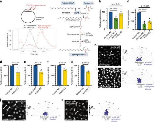
Wnt5b signaling regulates the levels of sphingolipids by modulating SPT activity.a Scheme of metabolic tracer assay. One-cell stage embryos were injected with (13C315N)-l-serine (heavy serine/serine*), which is incorporated into 3-ketosphinganine by SPT, losing one heavy carbon. Incorporation of the remaining three heavy carbons into the downstream sphingoid base sphingosine is used as a direct in vivo readout for SPT activity. Heavy sphingosine (red) co-elutes with sphingosine (black), but it can be distinguished due to its higher mass/charge ratio (m/z). These experiments were done three times independently for each condition. b, c Relative amounts of heavy sphingosine in wnt5a morphant embryos (b), wnt5b morphants (c) or embryos treated with Myriocin (b, c) from three independent experiments. d, e Relative amounts of C18 sphinganine (d) or C18 sphingosine (e) in wnt5a morphant embryos from three independent experiments. f, g Relative amounts of C18 sphinganine (f) or C18 sphingosine (g) in wnt5b morphant embryos from three independent experiments. Unpaired, two-tailed student’s t-test was used in (b–g). All error bars represent the standard error of the mean (SEM). h–k Orientation of division of epiblast cells with respect to the A/V embryonic axis determined by using H2A.F/Z:GFP transgenic line to monitor chromosome separation during anaphase. Left, representative confocal images of dorsal epiblast cells expressing H2A-GFP in wnt5a morphants (h), in wnt5a morphants treated with sphinganine (0.5 nl of 10 μM) (i), in wnt5b morphants (j) and in wnt5b morphants treated with sphinganine (0.5 nl of 25 μM) (k). Division axes are marked by arrows. Animal pole is up. Scale bars: 10 µm. Right, polar graphs showing the frequency distribution of the angle between the division axis and the A/V embryonic axis in wnt5a morphants (h) and in wnt5a morphants treated with sphinganine (0.5 nl of 10 μM) (i), wnt5b morphants (j) and in wnt5b morphants treated with sphinganine (0.5 nl of 25 μM) (k). n (number of cell divisions analyzed) over N (number of embryos). Note that rescue of randomization of division in wnt5b morphants requires higher amounts of sphinganine than for wnt5a morphants. In (h–k), χ2 test was used (see Supplementary Table 1). Source data are provided as a Source Data file.
|