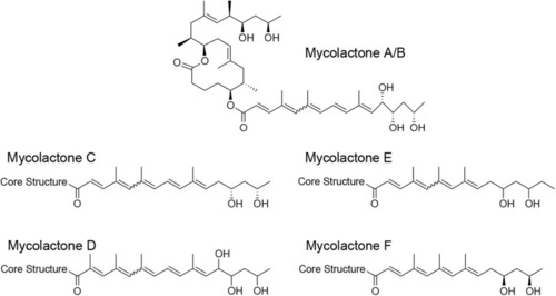
Structure of mycolactone variants. Mycolactone congeners were shown to be produced by M. ulcerans strains isolated from BU patients in Africa (mycolactone A/B), Australia (mycolactone C), and China (mycolactone D) and from fish and frogs of different geographical origin (mycolactone E and F).

Structure and cellular composition of granulomas. Early innate M. marinum granulomas, as described in zebrafish embryos, are composed of compact, organized aggregates of macrophages that have either transformed into epithelioid cells with tightly interdigitated cell membranes or have fused to become multinucleated giant cells. Bacilli are found both extracellularly in the necrotic core of the granuloma and intracellularly within phagocytes (A). The characteristic mature tuberculous granuloma is populated by many other cell types such as neutrophils, dendritic cells, B and T cells, natural killer cells, and foam cells, which are macrophages with accumulated lipids (B) (created with biorender.com).

Early host immune response to M. ulcerans infection illustrated in histological sections of a nodular lesion 6 weeks after subcutaneous infection of pigs (A–D) (from Bolz et al., 2016b) and in a sketch depicting the cellular composition of the lesion (E) (created with biorender.com). Histopathological analysis of pig lesions shows a central necrotic core containing extracellular M. ulcerans bacilli (A1,A2; AFB stained with Ziehl–Neelsen staining) and neutrophilic debris (B1,B2). The necrotic core is surrounded by layers of neutrophils (B1,B2) and macrophages (C1,C2), heavily interspersed with T-cells (D1,D2).

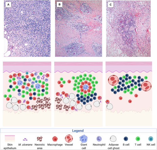
Immune reconstitution responses after chemotherapy illustrated in histological sections of a human BU lesion (from Schütte et al., 2007) and in a sketch of the participating immune components (created with biorender.com). Three main types of cellular infiltration have been observed in BU patients after treatment with the standard antibiotic regimen, including diffuse, heterogeneous cellular infiltration of the connective and adipose tissue (100× magnified) (A), granuloma-like structures in the connective tissue (40× magnified) (B), and follicle-like lymphocyte structures adjacent to vessels (40× magnified) (C).

Acknowledgments
This image is the copyrighted work of the attributed author or publisher, and ZFIN has permission only to display this image to its users. Additional permissions should be obtained from the applicable author or publisher of the image. Full text @ Front Microbiol