
FIGURE
FIGURE 3
- ID
- ZDB-FIG-200613-9
- Publication
- Röltgen et al., 2020 - Buruli ulcer: The Efficacy of Innate Immune Defense May Be a Key Determinant for the Outcome of Infection With Mycobacterium ulcerans
- Other Figures
- All Figure Page
- Back to All Figure Page
FIGURE 3
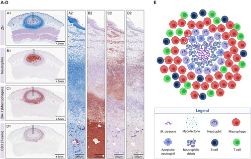
Early host immune response to |
Expression Data
Expression Detail
Antibody Labeling
Phenotype Data
Phenotype Detail
Acknowledgments
This image is the copyrighted work of the attributed author or publisher, and
ZFIN has permission only to display this image to its users.
Additional permissions should be obtained from the applicable author or publisher of the image.
Full text @ Front Microbiol