- Title
-
Expression of three P4- phospholipid Flippases-atp11a, atp11b, and atp11c in zebrafish (Danio rerio)
- Authors
- Hawkey-Noble, A., Umali, J., Fowler, G., French, C.R.
- Source
- Full text @ Gene Expr. Patterns
|
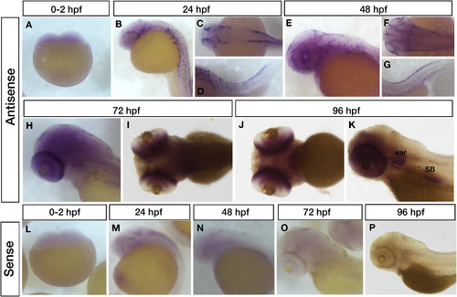
Spatial and temporal expression of zebrafish atp11a in wild-type embryos. Gene specific atp11a expression (antisense) at 0–2 hpf (2–4 cells; lateral view) demonstrating a lack of maternal deposition (A), 24 hpf (B: lateral, C: dorsal; and D: lateral tail) and 48 hpf (E: lateral, F: dorsal; and G: lateral tail) showing staining consistent with neural crest cell expression, 72 hpf (H: dorsolateral and I: dorsal) and 96 hpf (J: ventral and K: lateral) with ubiquitous expression in the head and high expression levels in both the developing eye and ear. Staining is also observed in the developing swim bladder (SB) at 96 (K). Non-specific staining (sense) is shown at the same timepoints for comparison (L–P). |
|
Detailed analysis of the zebrafish eye showing specific atp11a expression in the photoreceptors, retinal pigment epithelium, and ciliary marginal zone. (A and B) atp11a expression observed in cryosections at 72 hpf. (C and D) atp11a expression at 96 hpf. (E) antisense and (F) sense probes of flat mount in situ hybridization of excised zebrafish eyes at the 72 hpf stage, further highlight atp11a expression in either the RPE and/or OPR while also illustrating expression in the CMZ. The lens and optic nerve (ON) are indicated for reference. RPE (retinal pigment epithelium), OPR (outer segment photoreceptor layer), IPR (Inner segment photoreceptor layer), CMZ (ciliary marginal zone). EXPRESSION / LABELING:
|
|
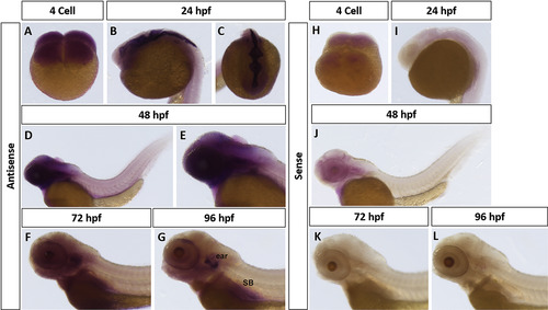
Spatial and temporal expression of zebrafish atp11b in wild-type embryos. Gene specific atp11b expression (antisense) at 0–2 hpf (4 cells) illustrating maternal deposition (A: lateral) and at 24 hpf (B: lateral and C: dorsal head) in the ventricle epithelial lining. Expression of atp11 b at 48 hpf (D and E; lateral) is ubiquitous in the head with concentrated expression in the brain tissue behind the eye and possibly within the eye itself. 72 hpf (F: lateral) and 96 hpf (G: lateral) expression is found in the ear and swim bladder. Non-specific staining (sense) is shown at the same timepoints for comparison (H–L). |
|
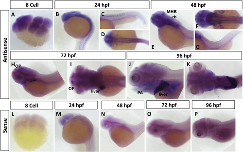
Spatial and temporal expression of zebrafish atp11c in wild-type embryos. Gene specific atp11c expression (antisense) at the 8-cell stage showing maternal deposition (A), 24 hpf (B: lateral, C: lateral tail, and D: dorsal) and 48 hpf (E: lateral, F: dorsal, G: lateral tail) showing expression in the mid-hindbrain boundary (MHB), anterior rhombomeres (rh), pharyngeal arches (PA), pectoral fin buds, olfactory pits (OP) and in a neuronal pattern along the tail. At 72 hpf (H: lateral and I: dorsal) and 96 hpf (J: lateral and K: ventral) there is ubiquitous expression in the head with increased expression in the jaw and OP. Both time points show a specific expression of atp11c in the liver. Non-specific staining (sense) is shown at the same timepoints for comparison (L–P). |
Reprinted from Gene expression patterns : GEP, 36, Hawkey-Noble, A., Umali, J., Fowler, G., French, C.R., Expression of three P4- phospholipid Flippases-atp11a, atp11b, and atp11c in zebrafish (Danio rerio), 119115, Copyright (2020) with permission from Elsevier. Full text @ Gene Expr. Patterns


