FIGURE SUMMARY
Title

Low doses of ionizing radiation enhance angiogenesis and consequently accelerate post-embryonic development but not regeneration in zebrafish

Authors
Marques, F.G., Carvalho, L., Sousa, J.S., Rino, J., Diegues, I., Poli, E., Pina, F., Saúde, L., Constantino Rosa Santos, S.
Source
Full text @ Sci. Rep.

LDIR upregulate the expression of several pro-angiogenic factors in Fli1:EGFP zebrafish in a VEGFR-dependent manner. Fli1:EGFP zebrafish larvae were exposed or not to 0.5 Gy at 3, 4 and 5 dpf and pre-treated or not with PTK/ZK, 30 minutes before each irradiation. The mRNA expression of flt1, kdr, angpt2a, tgfb2, fgf2, and cyr61 from whole cell suspension and endothelial sorter cells was quantified by qRT-PCR and normalized to elongation factor 1. (A) Data represent the relative mRNA levels of whole cell suspension and endothelial sorted cells from larvae exposed LDIR relative to the non-irradiated one (dashed line), in triplicate measurements. **P < 0.01; ***P < 0.001; ns, non-significant. (B) Data represent the fold change in mRNA expression of whole cell suspension and endothelial sorted cells of irradiated larvae exposed to PTK-ZK relative to non-irradiated and untreated ones, in triplicate measurements. Data are representative of six independent experiments.

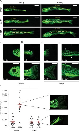
LDIR accelerate zebrafish development in a VEGFR-dependent manner. Fli1:EGFP zebrafish larvae were exposed or not to 0.5 Gy at 3, 4 and 5 dpf, pre-treated or not with PTK/ZK, 30 minutes before each irradiation and photographed over-time. (A) Representative images of the vasculature from non-irradiated and irradiated zebrafish at the 27th, 33rd and 38th dpf. The standard length (SL), in mm, was measured at each time-point for irradiated and non-irradiated zebrafish. (B–D) Post-embryonic development progress indicators were assessed at the 27th and 33rd dpf: (B) Head shape; (C) notochord flexion; (D) caudal fin; and (E) anal and dorsal fin. and Scale bars, 1 mm (A), 500 μm (C,D). (F) At the 33rd dpf, developmental stage was established by quantification of vascular caudal fin area, using ImageJ. Representative images of the median phenotypes are showed next to the graph. Scale bars, 500 μm. Data are represented as mean ± SEM and two-way ANOVA test was used to determine differences between experimental groups; ***P < 0.001.

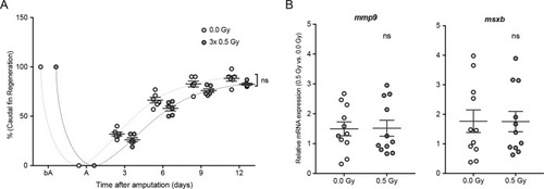
Wound healing and blastema formation, triggering the regeneration process, are not affected by LDIR after amputation in zebrafish. The caudal fin of adult Fli1:EGFP zebrafish was amputated at mid-fin level and immediately exposed or not to 0.5 Gy for 3 consecutive days. (A) Caudal fin regeneration was followed over time, and regenerated area was quantified (in pixels) using ImageJ software. Data are represented as mean ± SEM and two-way with repeated measures ANOVA test was used to determine differences between experimental groups; ns, non-significant. (B) The mRNA expression levels of mmp9 (wound-healing marker) and msxb (blastema formation marker) were determined by qRT-PCR and normalized to elongation factor 1. Data represent the relative mRNA expression in irradiated tissue compared to non-irradiated ones. Data are represented as mean ± SEM of 12 independent zebrafish and differences between groups was assessed by independent samples T-test; ns, non-significant.

Acknowledgments
This image is the copyrighted work of the attributed author or publisher, and ZFIN has permission only to display this image to its users. Additional permissions should be obtained from the applicable author or publisher of the image. Full text @ Sci. Rep.