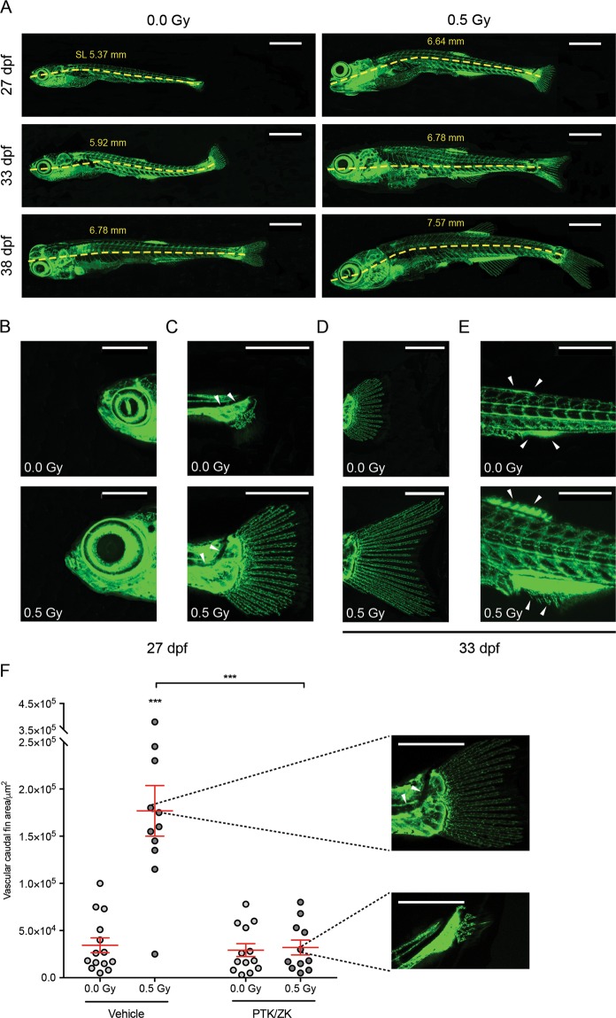
LDIR accelerate zebrafish development in a VEGFR-dependent manner. Fli1:EGFP zebrafish larvae were exposed or not to 0.5 Gy at 3, 4 and 5 dpf, pre-treated or not with PTK/ZK, 30 minutes before each irradiation and photographed over-time. (A) Representative images of the vasculature from non-irradiated and irradiated zebrafish at the 27th, 33rd and 38th dpf. The standard length (SL), in mm, was measured at each time-point for irradiated and non-irradiated zebrafish. (B–D) Post-embryonic development progress indicators were assessed at the 27th and 33rd dpf: (B) Head shape; (C) notochord flexion; (D) caudal fin; and (E) anal and dorsal fin. and Scale bars, 1 mm (A), 500 μm (C,D). (F) At the 33rd dpf, developmental stage was established by quantification of vascular caudal fin area, using ImageJ. Representative images of the median phenotypes are showed next to the graph. Scale bars, 500 μm. Data are represented as mean ± SEM and two-way ANOVA test was used to determine differences between experimental groups; ***P < 0.001.
Acknowledgments
This image is the copyrighted work of the attributed author or publisher, and
ZFIN has permission only to display this image to its users.
Additional permissions should be obtained from the applicable author or publisher of the image.
Full text @ Sci. Rep.
Your Input Welcome
Thank you for submitting comments. Your input has been emailed to ZFIN curators who may contact you if
additional information is required.
Oops. Something went wrong. Please try again later.