FIGURE SUMMARY
Title

Interference with the Cannabinoid Receptor CB1R Results in Miswiring of GnRH3 and AgRP1 Axons in Zebrafish Embryos

Authors
Zuccarini, G., D'Atri, I., Cottone, E., Mackie, K., Shainer, I., Gothilf, Y., Provero, P., Bovolin, P., Merlo, G.R.
Source
Full text @ Int. J. Mol. Sci.

Survival and hatching rates in zebrafish embryos treated with CB1R ligands. (A) Hatching rate (panels on the left) and survival rate (panels on the right) of gnrh3::EGFP zebrafish embryos following treatments with WIN55, Rimonabant or AM251, at the indicated doses, examined at the indicated embryonic age. Results are expressed as % over the total number of embryos examined. (B) Same as in (A), but using agrp1::mCherry embryos. *** = p < 0.001.

Survival and hatching rates in zebrafish embryos treated with CB1R ligands. (A) Hatching rate (panels on the left) and survival rate (panels on the right) of gnrh3::EGFP zebrafish embryos following treatments with WIN55, Rimonabant or AM251, at the indicated doses, examined at the indicated embryonic age. Results are expressed as % over the total number of embryos examined. (B) Same as in (A), but using agrp1::mCherry embryos. *** = p < 0.001.

Pharmacological manipulation of CB1R on GnRH3 neurons in zebrafish embryos. (A) Scheme showing the observation plane used for the micrographs in (C–F). (B) Low magnification of the head piece of gnrh3::EGFP embryos, viewed in combined fluorescence and bright field illumination. Red arrows indicate the EGFP+ cells associated to the terminal nerve. (CF) Representative images of EGFP+ neurons and their projections present in the nasal and basal forebrain regions of embryos at 72 hpf, treated either with DMSO only (negative control; C), with WIN55 (D), Rimonabant (E) or AM251 (F), at the indicated doses. Scale bar is reported in D. White arrows indicate misguided EGFP+ projections, and white asterisks indicate altered organization of EGFP+ fibers at the anterior commissure. (G) Bright-field low magnification images of whole embryos treated with DMSO only, WIN55, Rimonabant or AM251, showing a normal general morphology and growth. (H) Quantification of the number of EGFP+ neurons in the nasal region of fish embryos treated with DMSO only (black bar), or with WIN55, Rimonabant or AM251 at the indicated doses. The color code is the same as in Figure 1. No significant difference in the number of EGFP+ neurons was observed following these treatments. (I–K) Quantification of the misguidance and altered commissural phenotypes, expressed as % of the number of embryos presenting the phenotype, upon treatment with WIN55 (I), Rimonabant (J) or AM251 (K). Color code as in Figure 1. Data are expressed as means ± SEM from three independent experiments. * = p < 0.05; ** = p < 0.01; *** = p < 0.001. AC, anterior commissure.

Effects of MO-mediated CB1R knockdown on GnRH3 neurons in zebrafish embryos. (A) Western blot analyses of protein extracts from uninjected or z-cb1r MO-injected zebrafish embryos. A significant depletion of the CB1R protein was detected in the treated embryos. (B) Real-time qPCR analyses of z-cb1r and three z-hox genes on RNA samples extracted from uninjected (open bars) or z-cb1r MO-injected (solid black bars) embryos. Expression is shown relative to the expression of the housekeeping gene mRNA (z-β-actin). The abundance of the control RNA is set as 1. A depletion of the z-cb1r mRNA in embryos injected with z-cb1r MO is observed, as compared to control ones. No change in the expression of z-hox genes was observed, indicating unaltered developmental progression. (CE) Representative images of EGFP GnRH3 neurons and projections in the nasal and the basal forebrain regions of zebrafish embryos, at the age 72 hpf. The observation plane is the same as in Figure 2. Scale bar is reported in C. Red arrows indicate EGFP+ neurons, white arrows indicate altered EGFP+ projections, and white asterisks indicate altered organization and fasciculation at the anterior commissure. Below each micrograph, the bright-field images show the general morphology and growth of the corresponding whole animal, with no difference between the three conditions. (F) Quantification of the number of EGFP+ neurons in the nasal region of uninjected (open bars), scrmbl MO-injected (solid grey bars) or z-cb1r MO-injected (solid black bars) embryos. A significant decrease was observed in embryos injected with the z-cb1r MO, compared to control ones. (G) Quantification of the misguidance and altered commissural phenotype, expressed as % of the number of embryos observed. A significant increase was observed in embryos injected with the z-cb1r MO, compared to control ones. Data are expressed as means ± SEM from three independent experiments. ** = p < 0.01; *** = p < 0.001. AC, anterior commissure.

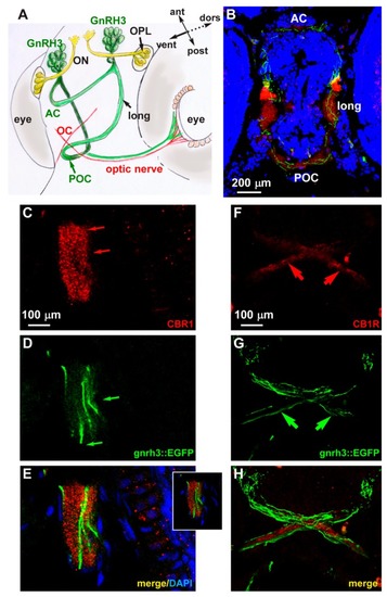
Expression of CB1R in developing zebrafish brain. (A) Scheme illustrating the position of the GnRH3 neurons relative to the position and orientation of the main forebrain commissure in the zebrafish brain (redrawn from [52]). (B) Low magnification double-fluorescent images of GnRH3::EGFP neurons (green) and IFL with an anti-CB1R antibody (red) on cryostatic sections of the zebrafish head at the age 72 hpf. (C–E) Higher magnification double-fluorescence images of the anterior commissure, showing overlapping localization of CB1R punctate staining (red) with EGFP+ fibers (green). Sections were counterstained with DAPI (blue). The merged signal is shown in E. Inset in E shows a single stack of the same image. Arrows indicate the fluorescence signal. (FH) Higher magnification double-fluorescence images of the optic chiasm, immunostained for CB1R (red), as in panels CE. Adjacent but non-overlapping expression was observed in these fibers. Arrows indicate the fluorescence signal. Scale bars are reported in panels B, in C (for C,D) and in F (for FH). AC, anterior commissure; ant, anterior; dors, dorsal; long, longitudinal tract; OC, optic chiasm; ON, olfactory nerve; OPL, olfactory placode; POC, postoptic commissure; post, posterior; RGC, retinal ganglion cells; ven, ventral.

EXPRESSION / LABELING:
Genes:
Fish:
Anatomical Terms:
Stage: Protruding-mouth

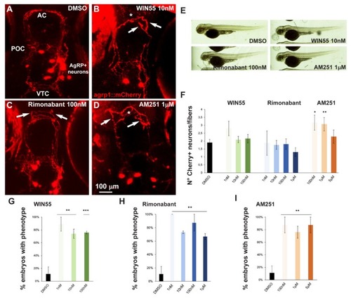
Effect of pharmacological manipulation of CB1R on AgRP1 neurons in zebrafish embryos. (A–D) Representative images of mCherry+ neurons and projections in the basal forebrain of embryos, at the age 96 hpf, treated with DMSO only as control (A), WIN55 (B), Rimonabant (C) or AM251 (D) at the indicated doses. The observation plane is the same as in Figure 2. Scale bar is reported in (D). White asterisks indicate altered fasciculation at the anterior commissure, and white arrows indicate misguided mCherry+ projections. (E) Bright-field, low-magnification images of whole fish embryos treated with DMSO only or with the indicated ligands. A normal general morphology and growth was observed. (F) Quantification of the ratio of mCherry+ neurons/mCherry+ fibers in embryos treated with DMSO only (solid black bar) or treated with WIN55, Rimonabant or AM251 at the indicated doses. Color code is the same as in Figure 1. Treatment with AM251 resulted in a higher neurons/fibers ratio. (G–I) Quantification of the misguidance and altered commissural phenotype, expressed as % of the number of embryos observed upon treatment with WIN55 (G), Rimonabant (H) or AM251 (I). Color code as in Figure 1. Data are expressed as means ± SEM from three independent experiments. * = p < 0.05; ** = p < 0.01; *** = p < 0.001. AC, anterior commissure; POC, postoptic commissure; VTC, ventral tegmental commissure.

Expression of z-stmn2a and z-stmn2b upon treatment with CB1R ligands or CB1R knockdown in early zebrafish embryos. (A) Real-time qPCR analyses of z-stmn2a (top histogram) and z-stmn2b (bottom histogram) on RNA samples obtained from embryos treated with the indicated CB1R ligands, at the indicated doses. Expression is shown relative to the expression of the housekeeping mRNA (z-β-actin). The abundance of the control RNA (DMSO) was set to 1. A reduced abundance of z-stmn2a mRNA was observed in embryos treated with the ligands, as compared to embryos treated with DMSO only. (B) Same analysis as above, on RNA samples obtained from uninjected (open bars), scrambled MO-injected (solid grey bars) or z-cb1r MO-injected (solid black bars) embryos. A reduced expression of z-stmn2a and z-stmn2b mRNAs was observed in embryos injected with z-cb1r MO as compared to the controls. * = p < 0.05; ** = p < 0.01; *** = p<0.001.

Acknowledgments
This image is the copyrighted work of the attributed author or publisher, and ZFIN has permission only to display this image to its users. Additional permissions should be obtained from the applicable author or publisher of the image. Full text @ Int. J. Mol. Sci.