IMAGE

Figure 4

ID
ZDB-IMAGE-200210-36
Genes
Source
Figures for Zuccarini et al., 2019
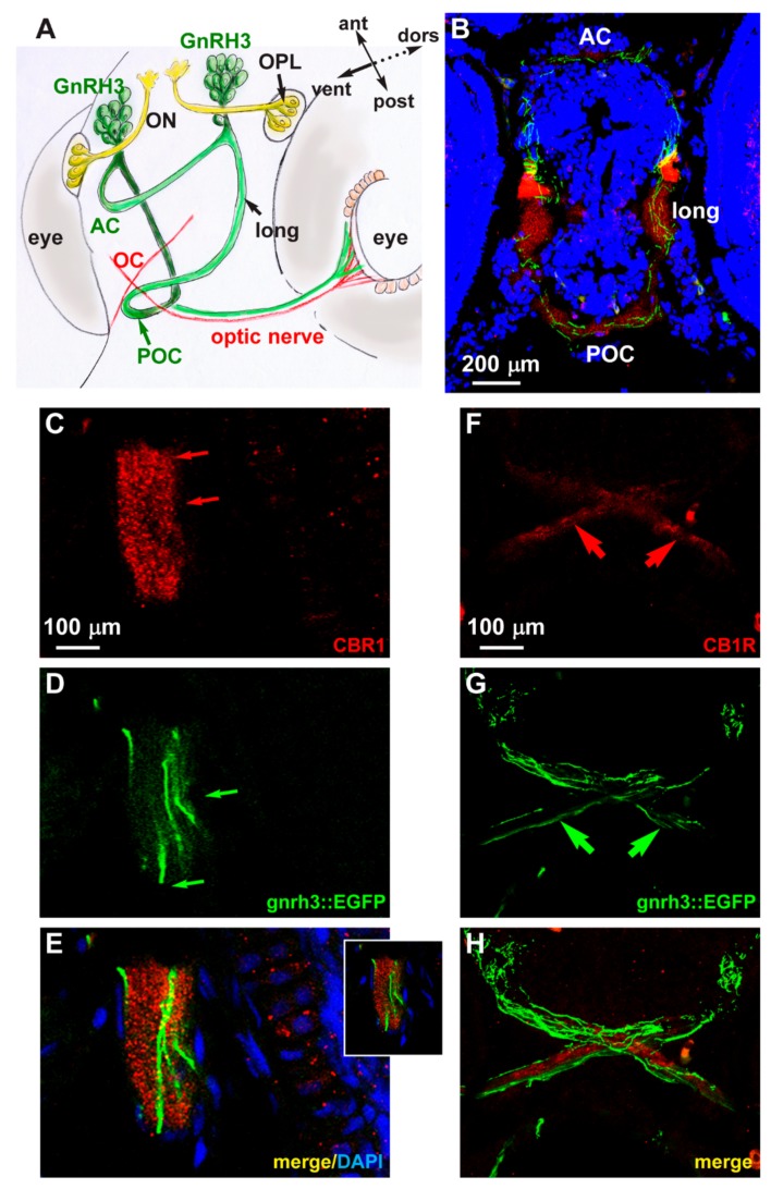
Image
Figure Caption

Figure 4

Expression of CB1R in developing zebrafish brain. (A) Scheme illustrating the position of the GnRH3 neurons relative to the position and orientation of the main forebrain commissure in the zebrafish brain (redrawn from [52]). (B) Low magnification double-fluorescent images of GnRH3::EGFP neurons (green) and IFL with an anti-CB1R antibody (red) on cryostatic sections of the zebrafish head at the age 72 hpf. (C–E) Higher magnification double-fluorescence images of the anterior commissure, showing overlapping localization of CB1R punctate staining (red) with EGFP+ fibers (green). Sections were counterstained with DAPI (blue). The merged signal is shown in E. Inset in E shows a single stack of the same image. Arrows indicate the fluorescence signal. (FH) Higher magnification double-fluorescence images of the optic chiasm, immunostained for CB1R (red), as in panels CE. Adjacent but non-overlapping expression was observed in these fibers. Arrows indicate the fluorescence signal. Scale bars are reported in panels B, in C (for C,D) and in F (for FH). AC, anterior commissure; ant, anterior; dors, dorsal; long, longitudinal tract; OC, optic chiasm; ON, olfactory nerve; OPL, olfactory placode; POC, postoptic commissure; post, posterior; RGC, retinal ganglion cells; ven, ventral.

Figure Data
Acknowledgments
This image is the copyrighted work of the attributed author or publisher, and ZFIN has permission only to display this image to its users. Additional permissions should be obtained from the applicable author or publisher of the image. Full text @ Int. J. Mol. Sci.