
- Title
-
Micro RNAs are involved in activation of epicardium during zebrafish heart regeneration
- Authors
- Ceci, M., Carlantoni, C., Missinato, M.A., Bonvissuto, D., Di Giacomo, B., Contu, R., Romano, N.
- Source
- Full text @ Cell Death Discov
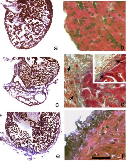
Histological staining using Masson’s trichrome on heart controls (a, b) and during regeneration (c–f). At 2 dpa (c) wide regenerative clot is clearly evidenced; in d a fibrous clot in blue is observed, while some undifferentiated cells that infiltrated the clot-surrounded epicardium are reactive (d, inset). We also notice some macrophages (d, inset). A 3 days (e) the clot is in resorption; in a magnification (f) epicardial stratification is observed. M myocytes, Ep epicardium (a bar = 1.5 mm; b bar = 45 μm; c, e bar = 1.5 mm; d, f bar = 45 μm) |
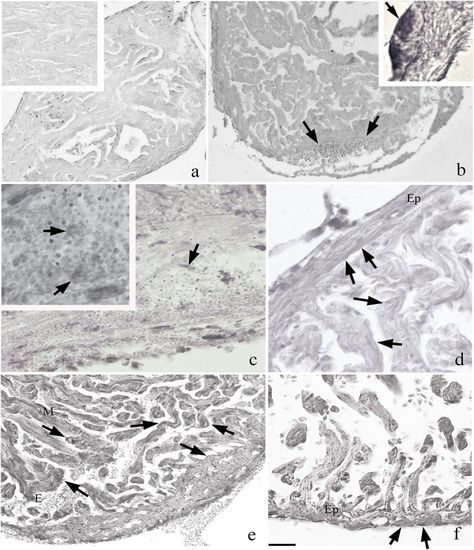
Immunohistochemistry with HSP70 on zebrafish heart. a HSP70 staining in the control. The reaction is marked in the samples of 2 and 3 dpa (b and d, respectively); in particular in the regions of the clot (c, and inset) and epicardium (Ep) (b, inset). In the region of cutting are visible at 2 dpa many rounded positive cells. From 14 days (f) onwards, the clot has almost disappeared and replaced by new myocytes (M); is visible to the evidenced several erythrocytes in the heart lumen (e). At days 30, (f) positivity is still reactive than in the control (a bar = 300 μm; inset = 75 μm; b bar = 300 μm and inset bar = 4 μm; c bar = 75 μm and inset bar = 45 μm; d bar = 150 μm and bar = 150 μm; f bar = 75 μm) |

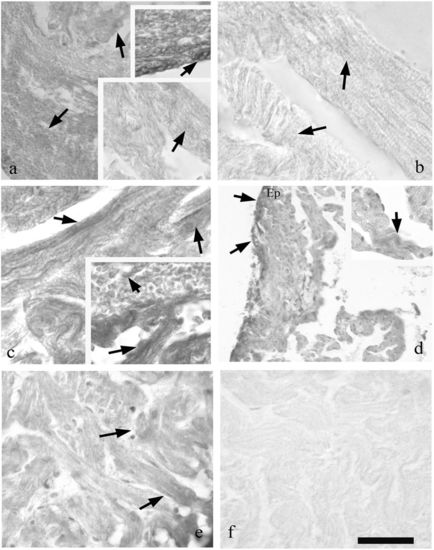
Immunohistochemistry with WT1 on regenerative heart (a bar = 300 µm; b bar = 150 µm; c bar = 75 µm; d bar = 45 µm; e bar = 50 µm; f bar = 45 µm; g bar = 150 µm). At 2 days, (a) there is a strong reactivity in the epicardium surrounding the organ. This positivity is present also in EPCs surrounding the clot (b) and in cells inside the clot (c). At 3 dpa, (d) the epicardium is reactive, but at 7 dpa (e) the signal in some epicardial portions is lower as compared with the parts next to the RC (f). In g, the control staining (bar = 75 µm). Ep epicardium |
Immunohistochemistry with cTnT on regenerating heart (a, e lower bar = 75 μm and upper inset bar = 45 μm; b bar = 54 μm; c bar = 45 μm and inset bar = 54 μm; d, bar = 150 μm and inset bar = 75 μm; and bar = 54 μm). At 2 days (a) are observed healthy muscle fibres with point reactivity (inset bottom) and highly reactive rounded cells (upper inset) among the heatly fibres. At 3 (b) and 7 (c) dpa next to healthy fibres (M) cTnT+ cells are evidenced close the clot (c, inset). As is shown in 14 days, (d) the epicardium (Ep) is cTnT posititve; infiltrated cells in the clot are also positive. At 30 dpa, (e) muscle fibres are reformed in the region of the cut and the reactivity for cTnT is comparable to the control. In f is the sample stained with omission of cTnT that shows no reactivity |
|