
|
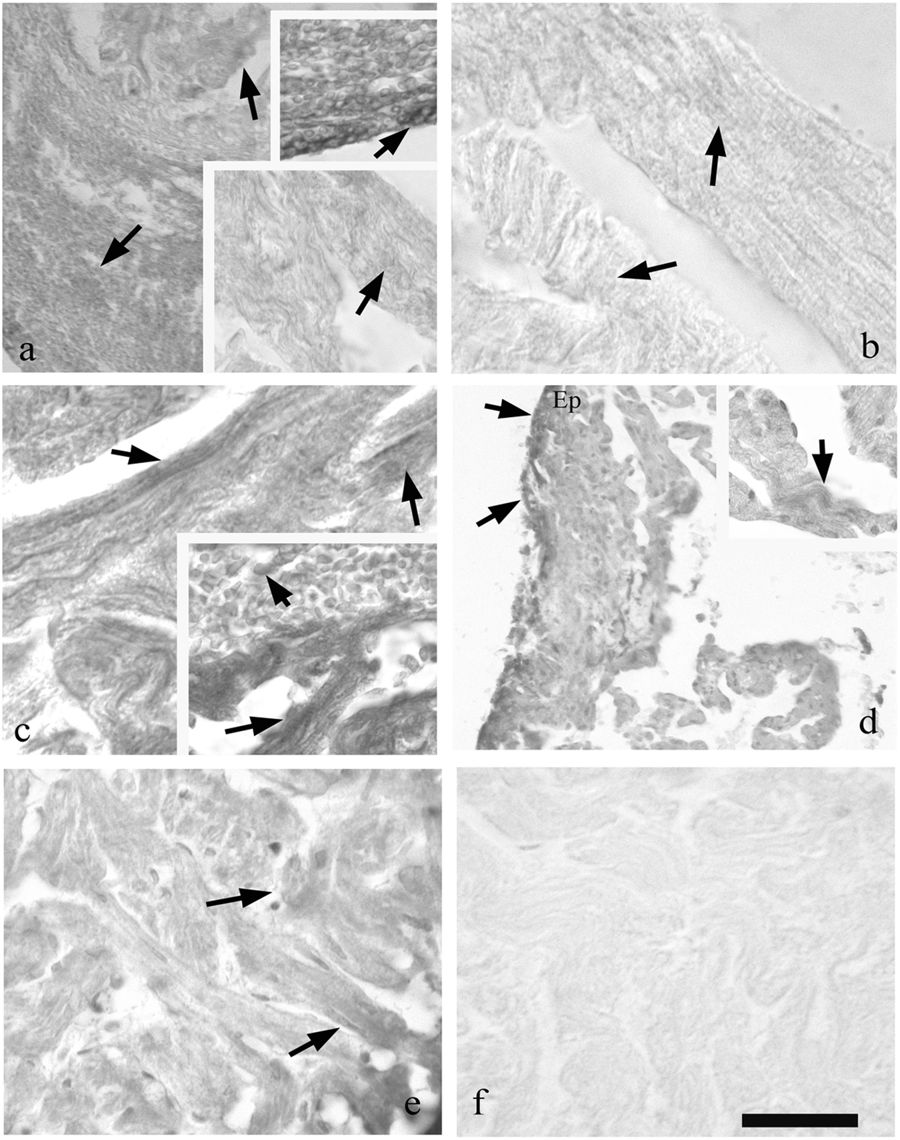
Fig. 6
Immunohistochemistry with cTnT on regenerating heart (a, e lower bar = 75 μm and upper inset bar = 45 μm; b bar = 54 μm; c bar = 45 μm and inset bar = 54 μm; d, bar = 150 μm and inset bar = 75 μm; and bar = 54 μm). At 2 days (a) are observed healthy muscle fibres with point reactivity (inset bottom) and highly reactive rounded cells (upper inset) among the heatly fibres. At 3 (b) and 7 (c) dpa next to healthy fibres (M) cTnT+ cells are evidenced close the clot (c, inset). As is shown in 14 days, (d) the epicardium (Ep) is cTnT posititve; infiltrated cells in the clot are also positive. At 30 dpa, (e) muscle fibres are reformed in the region of the cut and the reactivity for cTnT is comparable to the control. In f is the sample stained with omission of cTnT that shows no reactivity