
FIGURE
Fig. 3
- ID
- ZDB-FIG-180629-11
- Publication
- Ceci et al., 2018 - Micro RNAs are involved in activation of epicardium during zebrafish heart regeneration
- Other Figures
- All Figure Page
- Back to All Figure Page
Fig. 3
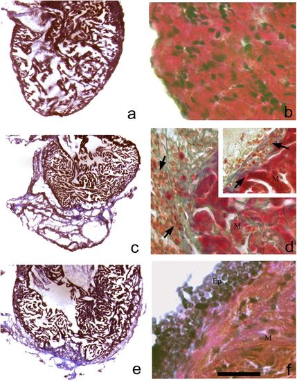
Histological staining using Masson’s trichrome on heart controls (a, b) and during regeneration (c–f). At 2 dpa (c) wide regenerative clot is clearly evidenced; in d a fibrous clot in blue is observed, while some undifferentiated cells that infiltrated the clot-surrounded epicardium are reactive (d, inset). We also notice some macrophages (d, inset). A 3 days (e) the clot is in resorption; in a magnification (f) epicardial stratification is observed. M myocytes, Ep epicardium (a bar = 1.5 mm; b bar = 45 μm; c, e bar = 1.5 mm; d, f bar = 45 μm) |
Expression Data
Expression Detail
Antibody Labeling
Phenotype Data
Phenotype Detail
Acknowledgments
This image is the copyrighted work of the attributed author or publisher, and
ZFIN has permission only to display this image to its users.
Additional permissions should be obtained from the applicable author or publisher of the image.
Full text @ Cell Death Discov