
- Title
-
Modelling acrylamide acute neurotoxicity in zebrafish larvae
- Authors
- Prats, E., Gómez-Canela, C., Ben-Lulu, S., Ziv, T., Padrós, F., Tornero, D., Garcia-Reyero, N., Tauler, R., Admon, A., Raldúa, D.
- Source
- Full text @ Sci. Rep.
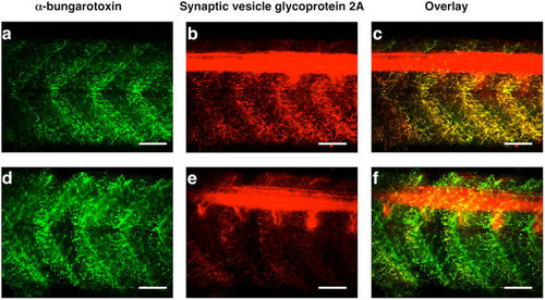
ACR reduces presynaptic nerve terminals in zebrafish larvae. At the neuromuscular junctions (NMJ) of the trunk, ACR-exposed larvae exhibit an strong reduction in the labelling of synaptic vesicle glycoprotein 2a (marker of synaptic terminals of the spinal motor neurons), whereas the α-bungarotoxin labelling (post-synaptic marker at the NMJ) labelling remains unaltered. Detail of the trunk, in lateral view, of control (a–c) and ACR-treated (d–f) larvae after co-labelling with α -bungarotoxin Alexa Fluor 488 conjugate (a,d) and SV2 antibody. The co-localization of the pre-synaptic and post-synaptic markers of NMJ is also showed (c,f). Scale bar: 100 μm. |
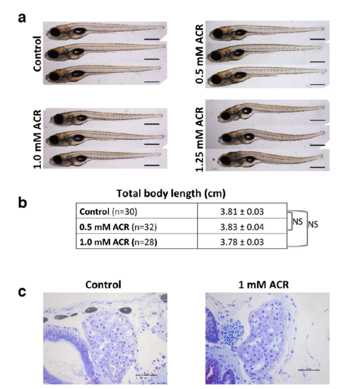
Determination of MTC for ACR systemic toxicity. (a) Gross morphology assessment shows that whereas 1.25 mM ACR is the LOEC, 1 mM ACR is the NOEC for this endpoint. (b) Morphometric analysis shows that 0.5-1.0 mM ACR has no effect on total length of the larvae. Statistical analysis was performed using one-way ANOVA with Dunnett’s multiple comparison test. Results represent mean ± sem; NS: not significant (c) Exposure to 1 mM ACR has no effect on liver morphology. Scale bar: (a) 500 m; (c) 50 m. |
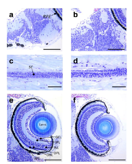
Histopathological analysis of control and 1 mM ACR exposed larvae. Representative semithin sections of the brain (a,b), spinal cord (c,d) and the retina (e,f) of control (a,c,e) and ACR-treated (b,d,f) zebrafish larvae. No histopathological changes were identified using this methodology. Abbreviations: GCL, ganglion cell layer; INL, inner nuclear layer; IPL, inner plexiform layer; ONL, outer nuclear layer; OPL; outer plexiform layer; RPE, retinal pigment epithelium; sc, spinal cord. Scale bar: (a,b,e,f) 100 μm, (c,d) 50 μm. |
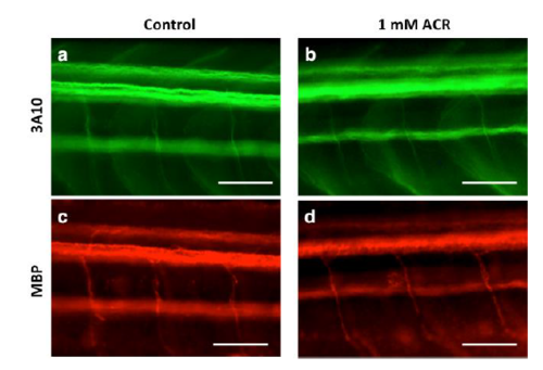
Axonal tracts and myelin sheets are not primary targets for acrylamide neurotoxicity in zebrafish larvae. Detail of the trunk, in lateral view, of control (a,c) and ACR-treated larvae (b,d) after double whole-mount immunofluorescence with 3A10 antibody labelling axonal tracts and MBP antibody labelling the myelin sheets. No clear differences in both structures were evident between the two groups. Scale bar: 100 m. |