
FIGURE
Fig. s5
- ID
- ZDB-FIG-180323-28
- Publication
- Prats et al., 2017 - Modelling acrylamide acute neurotoxicity in zebrafish larvae
- Other Figures
- All Figure Page
- Back to All Figure Page
Fig. s5
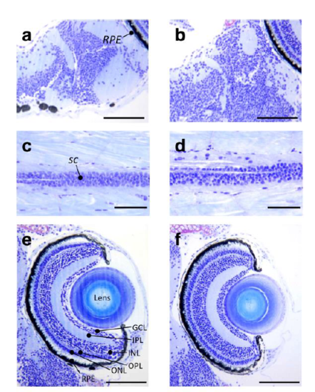
Histopathological analysis of control and 1 mM ACR exposed larvae. Representative semithin sections of the brain (a,b), spinal cord (c,d) and the retina (e,f) of control (a,c,e) and ACR-treated (b,d,f) zebrafish larvae. No histopathological changes were identified using this methodology. Abbreviations: GCL, ganglion cell layer; INL, inner nuclear layer; IPL, inner plexiform layer; ONL, outer nuclear layer; OPL; outer plexiform layer; RPE, retinal pigment epithelium; sc, spinal cord. Scale bar: (a,b,e,f) 100 μm, (c,d) 50 μm. |
Expression Data
Expression Detail
Antibody Labeling
Phenotype Data
Phenotype Detail
Acknowledgments
This image is the copyrighted work of the attributed author or publisher, and
ZFIN has permission only to display this image to its users.
Additional permissions should be obtained from the applicable author or publisher of the image.
Full text @ Sci. Rep.