
- Title
-
Beta-Catenin and Plakoglobin Expression during Zebrafish Tooth Development and Replacement
- Authors
- Verstraeten, B., van Hengel, J., Huysseune, A.
- Source
- Full text @ PLoS One
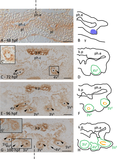
Plakoglobin distribution during the development of first-generation teeth. Cross-sections through the pharyngeal region of a 48 hpf (A), 72 hpf (C), 96 hpf (E) and 100 hpf (G) zebrafish embryo and corresponding schematic drawings in B, D, F and H. A,B: Initiation stage of tooth 4V1; the pharyngeal epithelium (ph.e.) expresses plakoglobin, in contrast to the mesenchymal cells. C,D: Morphogenesis stage of tooth 4V1; plakoglobin is clearly expressed at the cell borders of the epithelial-derived tissue (arrowhead). The keratinized pad (k.p.) opposite the developing teeth, strongly expresses plakoglobin. Boxed area in C is magnified in C’. E,F: Tooth 4V1 in late cytodifferentiation stage; teeth 3V1 and 5V1 in early cytodifferentiation stage. All teeth present display plakoglobin expression in epithelial-derived cell layers. G,H: Initiation of the first replacement tooth (arrow), 4V2. The epithelial outgrowth shows plakoglobin expression while the condensed mesenchyme is negative. Boxed area in G is magnified in G’. Diagrams: blue patch: placode; green line: contour of the tooth; orange: tooth matrix. Orientation: dorsal to the top, ventral to the bottom of each figure; dashed line indicates mediosagittal plane. Additional abbreviations: ph.c: pharyngeal cavity; pl: placode; *: dental papilla. Scale bars = 20 μm. |
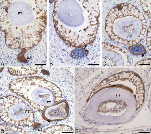
Plakoglobin distribution in adult replacement teeth. Cross-sections through one of the paired fifth branchial arches of an adult zebrafish. A: In contrast to the surrounding mesenchyme, all the cells constituting the successional lamina (s.l.) express plakoglobin at their cell membrane. Also the cells of the crypt epithelium express plakoglobin. B: Morphogenesis stage; the condensed mesenchyme constituting the dental papilla (black asterisk) is plakoglobin-negative. The enamel organ (arrowhead), including cervical loops (white asterisk), is strongly expressing plakoglobin. C: During early cytodifferentiation, the expression of plakoglobin remains limited to the inner and outer dental epithelium (arrowhead). D: Tooth in late cytodifferentiation stage, showing plakoglobin expression in the enamel organ (arrowhead) and in the differentiating odontoblasts lining the dental papilla (asterisk). E: Erupted functional tooth (FT) showing that even in a fully developed tooth plakoglobin expression persists in the reduced enamel organ (arrow). Orientation: dorsal to the top, ventral to the bottom, medial to the right and lateral to the left of the figure. Additional abbreviations: c: crypt surrounding the tip of the functional predecessor. Scale bars = 50 μm. EXPRESSION / LABELING:
|

Comparing tooth development in control-injected and plakoglobin morpholino-injected zebrafish. A,C,E: cross-sections through the pharyngeal cavity (ph.c.) of control-injected zebrafish at 72, 80 and 96 hpf, respectively; B,D,F: cross-sections at approximately the same level in plakoglobin morphant (MO) zebrafish at 72, 80 and 96 hpf, respectively. A,B: In both control-injected and morphant embryos the first tooth to develop, 4V1 is in early cytodifferentiation stage. C,D: Tooth 4V1 has continued to develop in control-injected and morphant embryos. Moreover, tooth 5V1 is also visible in both dentitions. E,F: The first three tooth positions develop normally in both control-injected and in morphant zebrafish. Orientation: dorsal to the top, ventral to the bottom of each figure; dashed line indicates mediosagittal plane. Additional abbreviations: ph.e: pharyneal epithelium, line: contour of the developing tooth. Scale bars = 20 μm. PHENOTYPE:
|
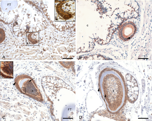
β-catenin distribution during the development of adult replacement teeth. A,A’: The successional lamina (s.l.) shows expression of β-catenin at the cell membrane and in some nuclei. The mesenchyme also displays nuclear β-catenin. B: β-catenin is detected only at the plasma membrane of the epithelial cells during morphogenesis stage (arrowhead). C: In the enamel organ (arrowhead) β-catenin is expressed at the plasma membrane of cells of both inner and outer dental epithelium. C’: The dental papilla (asterisk) shows odontoblasts with membrane-bound and nuclear β-catenin expression. D: During late cytodifferentiation stage, β-catenin remains expressed in both the inner and outer dental epithelium as well as in the polarized odontoblasts (arrow) adjoining the tooth matrix. Orientation: dorsal to the top, ventral to the bottom, medial to the right and lateral to the left of the figure. Additional abbreviations: c: crypt slightly posterior to the tip of the functional predecessor; FT: functional tooth. Scale bars = 50 µm. EXPRESSION / LABELING:
|