
Fig. 4
- ID
- ZDB-FIG-171108-37
- Publication
- Verstraeten et al., 2016 - Beta-Catenin and Plakoglobin Expression during Zebrafish Tooth Development and Replacement
- Other Figures
- All Figure Page
- Back to All Figure Page
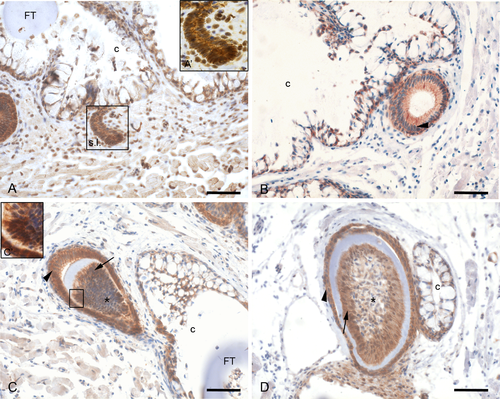
β-catenin distribution during the development of adult replacement teeth. A,A’: The successional lamina (s.l.) shows expression of β-catenin at the cell membrane and in some nuclei. The mesenchyme also displays nuclear β-catenin. B: β-catenin is detected only at the plasma membrane of the epithelial cells during morphogenesis stage (arrowhead). C: In the enamel organ (arrowhead) β-catenin is expressed at the plasma membrane of cells of both inner and outer dental epithelium. C’: The dental papilla (asterisk) shows odontoblasts with membrane-bound and nuclear β-catenin expression. D: During late cytodifferentiation stage, β-catenin remains expressed in both the inner and outer dental epithelium as well as in the polarized odontoblasts (arrow) adjoining the tooth matrix. Orientation: dorsal to the top, ventral to the bottom, medial to the right and lateral to the left of the figure. Additional abbreviations: c: crypt slightly posterior to the tip of the functional predecessor; FT: functional tooth. Scale bars = 50 µm. |
| Antibody: | |
|---|---|
| Fish: | |
| Anatomical Terms: | |
| Stage: | Adult |