
Fig. 2
- ID
- ZDB-IMAGE-171108-33
- Antibodies
- Publication
- Verstraeten et al., 2016 - Beta-Catenin and Plakoglobin Expression during Zebrafish Tooth Development and Replacement
- All Figures
- Figures for Verstraeten et al., 2016
|
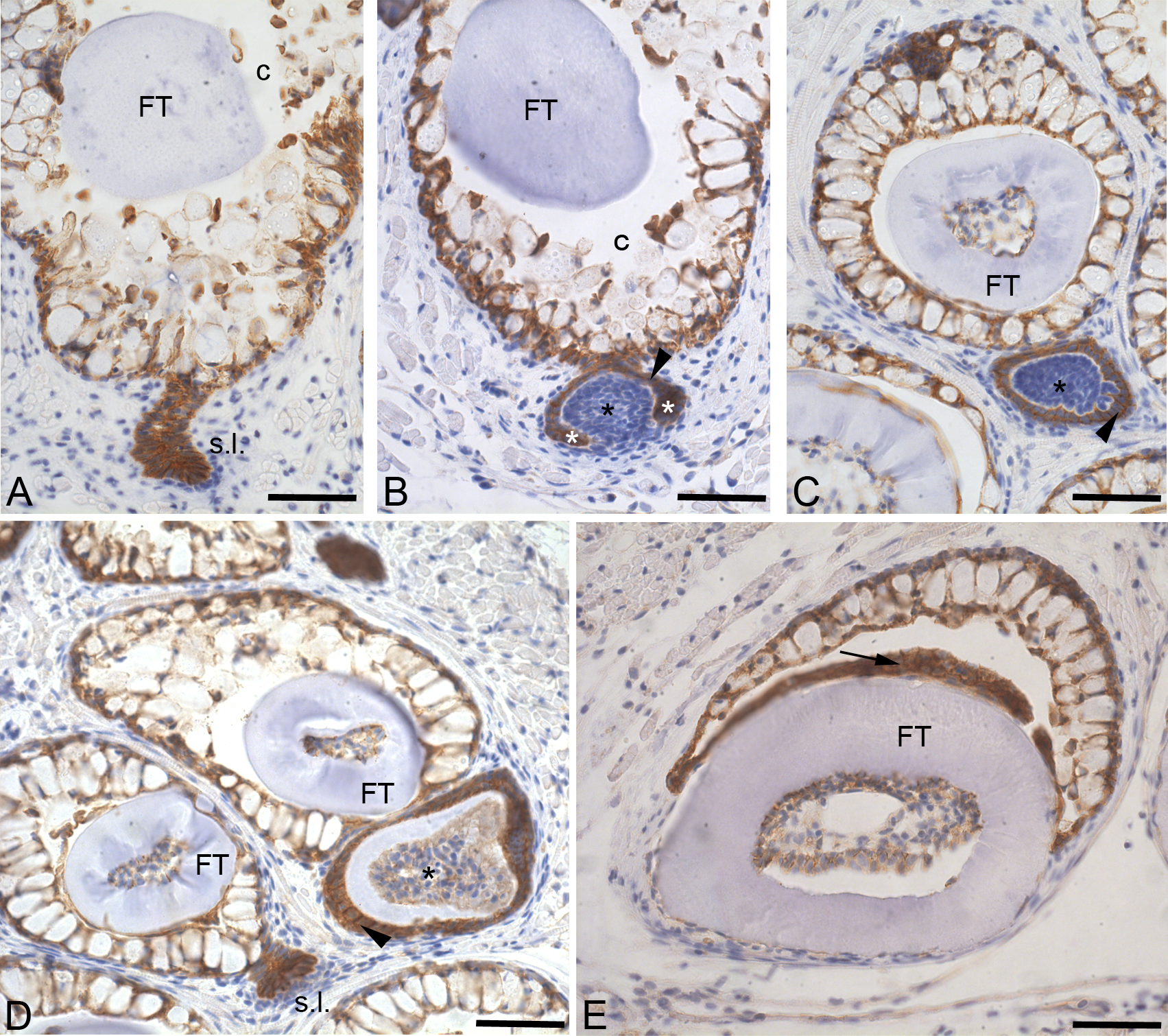
Fig. 2
Plakoglobin distribution in adult replacement teeth.
Cross-sections through one of the paired fifth branchial arches of an adult zebrafish. A: In contrast to the surrounding mesenchyme, all the cells constituting the successional lamina (s.l.) express plakoglobin at their cell membrane. Also the cells of the crypt epithelium express plakoglobin. B: Morphogenesis stage; the condensed mesenchyme constituting the dental papilla (black asterisk) is plakoglobin-negative. The enamel organ (arrowhead), including cervical loops (white asterisk), is strongly expressing plakoglobin. C: During early cytodifferentiation, the expression of plakoglobin remains limited to the inner and outer dental epithelium (arrowhead). D: Tooth in late cytodifferentiation stage, showing plakoglobin expression in the enamel organ (arrowhead) and in the differentiating odontoblasts lining the dental papilla (asterisk). E: Erupted functional tooth (FT) showing that even in a fully developed tooth plakoglobin expression persists in the reduced enamel organ (arrow). Orientation: dorsal to the top, ventral to the bottom, medial to the right and lateral to the left of the figure. Additional abbreviations: c: crypt surrounding the tip of the functional predecessor. Scale bars = 50 μm.