
Fig. 3
- ID
- ZDB-IMAGE-171108-34
- Publication
- Verstraeten et al., 2016 - Beta-Catenin and Plakoglobin Expression during Zebrafish Tooth Development and Replacement
- All Figures
- Figures for Verstraeten et al., 2016
|
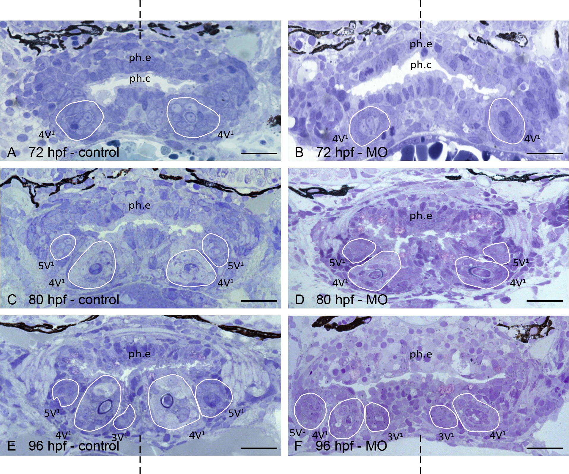
Fig. 3
Comparing tooth development in control-injected and plakoglobin morpholino-injected zebrafish.
A,C,E: cross-sections through the pharyngeal cavity (ph.c.) of control-injected zebrafish at 72, 80 and 96 hpf, respectively; B,D,F: cross-sections at approximately the same level in plakoglobin morphant (MO) zebrafish at 72, 80 and 96 hpf, respectively. A,B: In both control-injected and morphant embryos the first tooth to develop, 4V1 is in early cytodifferentiation stage. C,D: Tooth 4V1 has continued to develop in control-injected and morphant embryos. Moreover, tooth 5V1 is also visible in both dentitions. E,F: The first three tooth positions develop normally in both control-injected and in morphant zebrafish. Orientation: dorsal to the top, ventral to the bottom of each figure; dashed line indicates mediosagittal plane. Additional abbreviations: ph.e: pharyneal epithelium, line: contour of the developing tooth. Scale bars = 20 μm.