- Title
-
Attractant and repellent cues cooperate in guiding a subset of olfactory sensory axons to a well-defined protoglomerular target
- Authors
- Taku, A.A., Marcaccio, C.L., Ye, W., Krause, G.J., Raper, J.A.
- Source
- Full text @ Development
|
sema3d and netrin 1b have complementary expression patterns in the zebrafish OB at 36hpf. (A) Maximum intensity projection spanning 30µm through a 36hpf or111-7:IRES:GAL4:UAS:citrine embryo. Ventral view, anterior is up and medial is to the right. Axons are shown in green, sema3d mRNA in red and netrin 1b mRNA in blue. (B-B′′) A single optical section through the inset in A. (C,C′) Single optical sections through a 36hpf embryo. Frontal view, dorsal is up and medial is to the right. Sections are arranged from anterior (left) to posterior (right). The distance from each section to the anteriormost part of the telencephalon is denoted in bottom left. (D) Diagram of a 36hpf embryo in lateral view. sema3d is expressed in the anterior OB. Some netrin 1b is detected in the anterior bulb but it extends further posteriorly. or111-7 transgene-expressing axons are positioned posterior to sema3d expression but within the netrin 1b expression domain. or111-7 transgenic axons are not present in the anteriormost portion of the telencephalon. sema3d expression wraps around the edge of the olfactory pit and is also present between the OE and nascent OB. OE, olfactory epithelium; OB, olfactory bulb. |
|
or111-7 transgenic axons misproject to the DZ in sema3d mutants. (A,B) Single optical sections through 72hpf or111-7:IRES:GAL4;UAS:citrine larvae (frontal view). Axons are in green. Dorsal is up and medial is to the right. Propidium iodide (blue) labels cell bodies, revealing protoglomeruli as cell-free regions. (C,D) Maximum intensity projections of serial optical sections from the larvae shown in A,B. (A,C) Wild-type or111-7 transgenic axons project to the CZ and LG1. (B,D) In sema3d mutants, a subset of or111-7 transgenic axons inappropriately projects to the DZ. Scale bar: 25µm. (E) The percentage of OBs that have a labeled projection to the DZ protoglomerulus. sema3d homozygous mutants are compared with heterozygous and wild-type siblings. Statistical significance was estimated using two-tailed Fisher′s exact test (P<0.05*). Error bars represent s.e. of the sample proportion. CZ, central zone; DZ, dorsal zone; LG1, lateral protoglomerulus 1; OE, olfactory epithelium. PHENOTYPE:
|
|
Four zebrafish neuropilins are expressed in or111-7 transgene-labeled neurons during development. (A-D) Single optical sections through the OE of 36hpf embryos (frontal view). mRNA is in magenta. Subsets of or111-7 transgene-labeled neurons (green) express (A) nrp1a (N=56 pits/764 cells), (B) nrp1b (N=16 pits/191 cells), (C) nrp2a (N=29 pits/610 cells) and (D) nrp2b (N=26 pits/384 cells). Scale bar: 25µm. (E) The percentage of or111-7 transgene-expressing cells expressing a given neuropilin at 36hpf. Error bars represent s.d. EXPRESSION / LABELING:
|
|
Loss of nrp1a phenocopies sema3d mutants. (A-D) Single optical sections through 72hpf or111-7 transgenic larvae (frontal view). Axons are in green. Dorsal is up and medial is to the right. Propidium iodide (blue) labels cell bodies revealing protoglomeruli as cell-free regions. WT, wild type. Scale bar: 25µm. (E-G) Percentage of OBs displaying a projection to a particular protoglomerulus or all non-protoglomerular regions (other) is shown. Statistical significance was estimated using two-tailed Fisher′s exact tests (P<0.05*, P<0.01**, P<0.001***; ns, not significant). Homozygous mutants are compared with wild-type and heterozygous siblings. Error bars represent s.e. of the sample proportion. (B,E) A subset of or111-7 transgene-labeled axons misproject to the DZ and to non-protoglomerular areas in nrp1a mutants. (C,F) nrp1b mutants do not have increased mistargeting errors to the DZ but do have increased misprojections to LG2. (D,G) A subset of or111-7 transgene-labeled axons also inappropriately targets the DZ in nrp2b mutants. CZ, central zone; DZ, dorsal zone; LG1, lateral protoglomerulus 1; LG2, lateral protoglomerulus 2. |
|
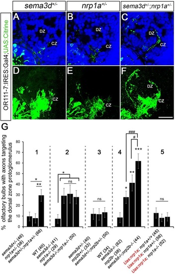
sema3d interacts genetically with nrp1a to promote axon targeting of or111-7 transgene-labeled axons to the CZ. (A-C) Single optical sections through 72hpf or111-7 transgenic larvae (frontal view). Axons are in green. Dorsal is up and medial is to the right. Propidium iodide (blue) labels cell bodies revealing protoglomeruli as cell-free regions. (D-F) Maximum intensity projections of serial optical sections from the larvae shown in A-C. Scale bar: 25µm. (G) The percentage of OBs with projections to the DZ protoglomerulus. Statistical significance was estimated using two-tailed (P<0.05*, P<0.01**, P<0.001***) or one-tailed (#P<0.05, ###P<0.001) Fisher′s exact tests. Error bars represent s.e. of the sample proportion. sema3d+/-;nrp1a+/- transheterozygotic larvae (C,F; 1 in G) have an increase in projections to the DZ compared with sema3d+/- heterozygotic (A,D) or nrp1a+/- heterozygotic (B,E) siblings. sema3d-/- mutants, nrp1a-/- mutants and sema3d-/-;nrp1a-/- double mutants have a similar increase in projections to the DZ as compared with wild-type siblings (2 in G). By contrast, sema3d+/-;nrp2b+/- transheterozygotes do not have an increase in projections to the DZ when compared with sema3d+/- heterozygotic or nrp2b+/- heterozygotic siblings (3 in G). sema3d-/-;nrp2b-/- double mutants have a greater frequency of projections to the DZ than sema3d-/- or nrp2b-/- siblings (4 in G). nrp1a-/- mutants expressing the UAS:nrp1a:UAS:citrine transgene do not have an increase in misprojections to the DZ or non-protoglomerular regions as compared with wild-type or heterozygotic siblings (5 in G). CZ, central zone; DZ, dorsal zone. PHENOTYPE:
|
|
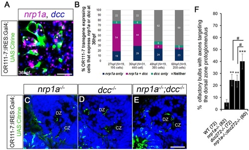
Relative contributions of Nrp1a and Dcc to DZ mistargeting. (A) A single optical section through a 36hpf or111-7 transgenic embryo (frontal view). Cell bodies are in green. Dorsal is up and medial is to the right. nrp1a mRNA is in magenta and dcc mRNA is in blue. (B) The percentages of or111-7-labeled neurons expressing nrp1a, dcc or both at 27, 36, 48 or 60hpf. (A,B) nrp1a and dcc are co-expressed in a large portion of or111-7-labeled neurons during early development (asterisks in A). (C-E) Single optical sections through 72hpf or111-7 transgenic larvae (frontal view). Axons are in green. Dorsal is up and medial is to the right. Propidium iodide (blue) labels cell bodies revealing protoglomeruli as cell-free regions. (F) The percentage of OBs with projections to the DZ protoglomerulus. (C-F) nrp1a-/- mutants, dcc-/- mutants and nrp1a-/-;dcc-/- double mutants have an increase in projections to the DZ as compared with wild-type siblings. nrp1a-/-;dcc-/- double mutants have a statistically significant increase in DZ errors when compared with single-mutant siblings. Statistical significance was estimated using two-tailed (**P<0.01, ***P<0.001) or one-tailed (#P<0.05) Fisher′s exact tests. Error bars represent s.e. of the sample proportion. CZ, central zone; DZ, dorsal zone. Scale bars: 10µm in A; 25µm in C-E. EXPRESSION / LABELING:
PHENOTYPE:
|
|
Sensory neurons expressing Omp or Trpc2 have no detectable targeting errors in sema3d mutants. A-B, Single optical sections through 72 hpf omp:RFP larvae (frontal view). Axons shown in red. Dorsal is up and medial is to the right of the image. C-D, Single optical sections through 72hpf trpc2:Venus larvae (frontal view). Axons shown in green. Propidium iodide (blue) labels cell bodies revealing protoglomeruli as cell-free regions. Scale bar (in D): A–D, 50µm. E-G, The percentage of olfactory bulbs displaying a projection to a particular protoglomerulus or non-protoglomerular regions (other) are shown. Homozygous sema3d mutants (black bars) are compared to heterozygous (grey bars) and wild-type (white bars) siblings. Statistical significance was estimated using two-tailed Fisher’s exact test (p < 0.05*, p < 0.01**, p < 0.001***). Error bars represent SE of the sample proportion. A, B, E, Omp positive axons project to the same protoglomeruli in sema3d mutants as in controls. C, D, F, The Trpc2:Venus projection is unaltered in sema3d mutants. G, OR111-7 transgene-expressing axons do not display an increase in projections to the listed protoglomeruli or to non-protoglomerular regions. H, single optical section through a 72 hpf sema3d+/-; nrp1a+/-;omp:RFP;OR111-7:IRES:GAL4;UAS:Citrine larva. The ectopic projection to DZ in sema3d+/-; nrp1a+/-;omp:RFP;OR111-7:IRES:GAL4;UAS:Citrine larvae is Omp positive. Scale bar (in H): H, 50µm. CZ, central zone; DZ, dorsal zone; MG, medial protoglomeruli; LG1, lateral protoglomerulus 1; LG2; lateral protoglomerulus 2, LG3, lateral protoglomerulus 3. |


