
Fig. S1
- ID
- ZDB-IMAGE-160218-22
- Publication
- Taku et al., 2016 - Attractant and repellent cues cooperate in guiding a subset of olfactory sensory axons to a well-defined protoglomerular target
- All Figures
- Figures for Taku et al., 2016
|
Fig. S1
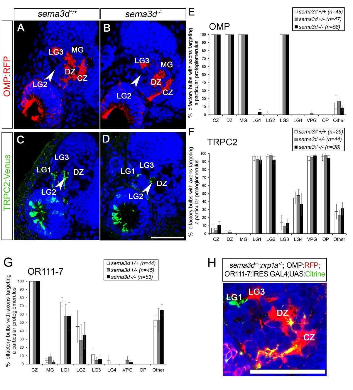
Sensory neurons expressing Omp or Trpc2 have no detectable targeting errors in sema3d mutants. A-B, Single optical sections through 72 hpf omp:RFP larvae (frontal view). Axons shown in red. Dorsal is up and medial is to the right of the image. C-D, Single optical sections through 72hpf trpc2:Venus larvae (frontal view). Axons shown in green. Propidium iodide (blue) labels cell bodies revealing protoglomeruli as cell-free regions. Scale bar (in D): A–D, 50µm. E-G, The percentage of olfactory bulbs displaying a projection to a particular protoglomerulus or non-protoglomerular regions (other) are shown. Homozygous sema3d mutants (black bars) are compared to heterozygous (grey bars) and wild-type (white bars) siblings. Statistical significance was estimated using two-tailed Fisher’s exact test (p < 0.05*, p < 0.01**, p < 0.001***). Error bars represent SE of the sample proportion. A, B, E, Omp positive axons project to the same protoglomeruli in sema3d mutants as in controls. C, D, F, The Trpc2:Venus projection is unaltered in sema3d mutants. G, OR111-7 transgene-expressing axons do not display an increase in projections to the listed protoglomeruli or to non-protoglomerular regions. H, single optical section through a 72 hpf sema3d+/-; nrp1a+/-;omp:RFP;OR111-7:IRES:GAL4;UAS:Citrine larva. The ectopic projection to DZ in sema3d+/-; nrp1a+/-;omp:RFP;OR111-7:IRES:GAL4;UAS:Citrine larvae is Omp positive. Scale bar (in H): H, 50µm. CZ, central zone; DZ, dorsal zone; MG, medial protoglomeruli; LG1, lateral protoglomerulus 1; LG2; lateral protoglomerulus 2, LG3, lateral protoglomerulus 3.