
- Title
-
Cis-regulatory control of human GLI2 expression in the developing neural tube and limb bud
- Authors
- Minhas, R., Pauls, S., Ali, S., Doglio, L., Khan, M.R., Elgar, G., Abbasi, A.A.
- Source
- Full text @ Dev. Dyn.

GLI2-associated CNEs upregulate GFP expression in live zebrafish embryos, using co-injection assays Intronic GLI2-associated conserved non-coding elements (CNE1-CNE5) mediate GFP expression pattern in live embryos at day-2 and day-3, fluorescent views (A-D), merged bright field and fluorescence views (E-J). Embryos A, C, E, G and J are ~26–33 hpf, while embryos B, D, F, H and I are ~48–54 hpf. Orientation of the embryos is anterior to left, dorsal to top, with lateral views. White arrowheads indicate GFP expressing cells. CNE1 drives GFP expression in muscle fibers in the trunk region (A), and in the neurons of the midbrain, hindbrain (B). CNE2 expresses GFP in the developing notochord at day-2 (~26–33 hpf) (C), as well as at day-3 (~48 hpf) (D). CNE3 drives GFP expression in the otic vesicle (E), and in the primary neurons of hindbrain (F). CNE4 showed GFP signal in muscle fibers at day-2 and day-3 (G & H). GFP expression was observed in the neurons of hindbrain (I), spinal cord and muscle fibers (J) by CNE5. hpf, hours post fertilization; mb, midbrain; hb, hindbrain; nc, notochord; sc, spinal cord; m, muscle; ov, otic vesicle. |
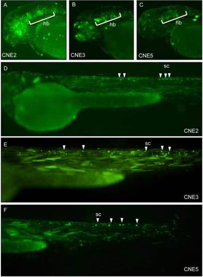
CNE2, CNE3, and CNE5 induced GFP expression was detected in various domains of CNS, using Tol2 based transgenic assays Patterns of GFP expression was observed by GLI2-associated CNEs in central nervous system. Images of live zebrafish embryos at ~48-54 hpf, lateral views, anterior to left, dorsal to top. Arrowheads and marked area point to GFP expressing cells. Using the Tol2 system, injected zebrafish embryos demonstrate that CNE2, CNE3 and CNE5 are central nervous system enhancers; induce GFP expression in primary neurons of hindbrain (A, B and C respectively) and spinal cord (D, E and F respectively). hb, hindbrain; sc, spinal cord. |
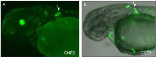
CNE2 and CNE5 are fin-specific enhancers Images of live zebrafish embryos at ~48-54 hpf, lateral views, anterior to left, dorsal to top. Arrowheads indicate GFP expressing cells. Using the Tol2 system, CNE2 (A) and CNE5 (B) induced GFP expression in the developing fin. f, fin. |

Duplicated copies of zebrafish CNE2 (CNE2a and CNE2b) induce overlapping GFP expression in hindbrain and pectoral fin GFP expression is presented in live embryos, by transiently transfected co-injection (A and B) and Tol2 transgenic assays (C, D, E, and F). All embryos were at ~48-54 hpf. Lateral views, anterior to the left, dorsal to the top. GFP expression is indicated by arrowheads and marked area in the following tissue or cell types: hindbrain (A and C) and pectoral fin (E) by Dr-gli2_CNE2a; hindbrain (B and D) and pectoral fin (F) by Dr-gli2_CNE2b. hb, hindbrain; f, fin. |