
|
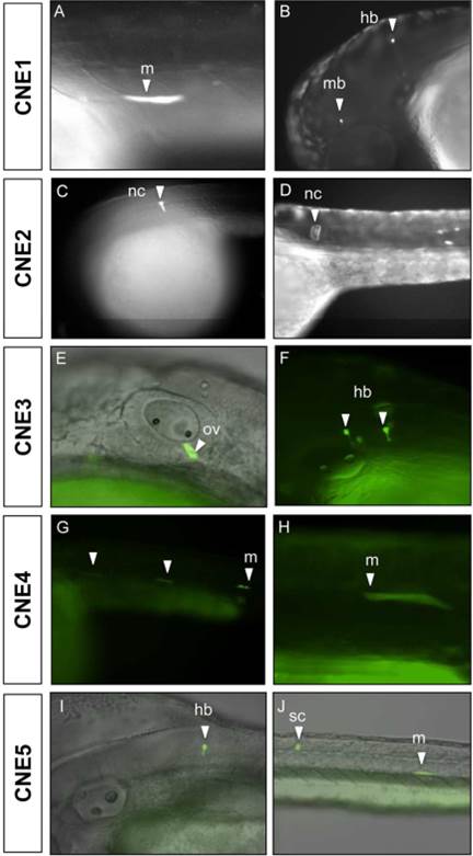
Fig. 3
GLI2-associated CNEs upregulate GFP expression in live zebrafish embryos, using co-injection assays
Intronic GLI2-associated conserved non-coding elements (CNE1-CNE5) mediate GFP expression pattern in live embryos at day-2 and day-3, fluorescent views (A-D), merged bright field and fluorescence views (E-J). Embryos A, C, E, G and J are ~26–33 hpf, while embryos B, D, F, H and I are ~48–54 hpf. Orientation of the embryos is anterior to left, dorsal to top, with lateral views. White arrowheads indicate GFP expressing cells. CNE1 drives GFP expression in muscle fibers in the trunk region (A), and in the neurons of the midbrain, hindbrain (B). CNE2 expresses GFP in the developing notochord at day-2 (~26–33 hpf) (C), as well as at day-3 (~48 hpf) (D). CNE3 drives GFP expression in the otic vesicle (E), and in the primary neurons of hindbrain (F). CNE4 showed GFP signal in muscle fibers at day-2 and day-3 (G & H). GFP expression was observed in the neurons of hindbrain (I), spinal cord and muscle fibers (J) by CNE5. hpf, hours post fertilization; mb, midbrain; hb, hindbrain; nc, notochord; sc, spinal cord; m, muscle; ov, otic vesicle.