
Image
|
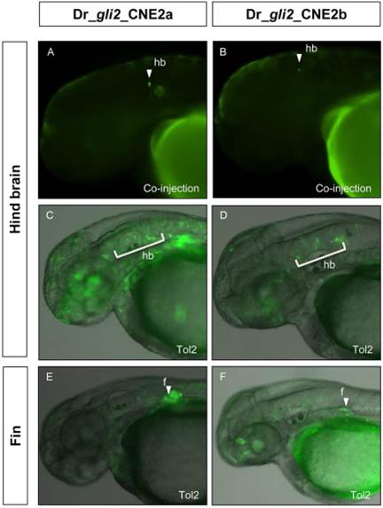
Figure Caption
Fig. 7
Duplicated copies of zebrafish CNE2 (CNE2a and CNE2b) induce overlapping GFP expression in hindbrain and pectoral fin
GFP expression is presented in live embryos, by transiently transfected co-injection (A and B) and Tol2 transgenic assays (C, D, E, and F). All embryos were at ~48-54 hpf. Lateral views, anterior to the left, dorsal to the top. GFP expression is indicated by arrowheads and marked area in the following tissue or cell types: hindbrain (A and C) and pectoral fin (E) by Dr-gli2_CNE2a; hindbrain (B and D) and pectoral fin (F) by Dr-gli2_CNE2b. hb, hindbrain; f, fin.
Acknowledgments
This image is the copyrighted work of the attributed author or publisher, and
ZFIN has permission only to display this image to its users.
Additional permissions should be obtained from the applicable author or publisher of the image.
Full text @ Dev. Dyn.