
|
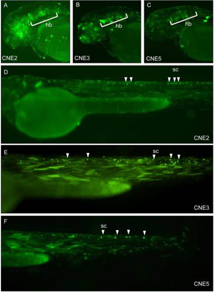
Fig. 4
CNE2, CNE3, and CNE5 induced GFP expression was detected in various domains of CNS, using Tol2 based transgenic assays
Patterns of GFP expression was observed by GLI2-associated CNEs in central nervous system. Images of live zebrafish embryos at ~48-54 hpf, lateral views, anterior to left, dorsal to top. Arrowheads and marked area point to GFP expressing cells. Using the Tol2 system, injected zebrafish embryos demonstrate that CNE2, CNE3 and CNE5 are central nervous system enhancers; induce GFP expression in primary neurons of hindbrain (A, B and C respectively) and spinal cord (D, E and F respectively). hb, hindbrain; sc, spinal cord.