
- Title
-
A Single Mutation in the Acetylcholine Receptor δ-Subunit Causes Distinct Effects in Two Types of Neuromuscular Synapses
- Authors
- Park, J.Y., Mott, M., Williams, T., Ikeda, H., Wen, H., Linhoff, M., Ono, F.
- Source
- Full text @ J. Neurosci.
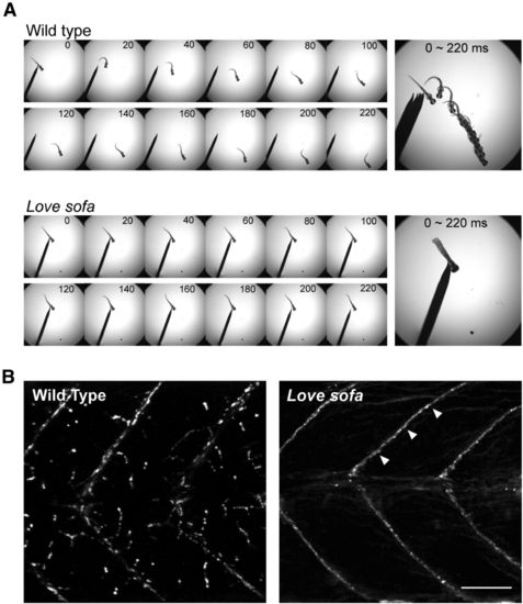
Phenotypes of the love sofa mutant. A, Time lapse images of a wild-type larva (top) and a love sofa mutant larva (bottom) at 2 dpf. Escape response was elicited by a gentle touch on the tail. Images at 0-220 ms after the start of the movement are shown with 20 ms intervals. B, Trunk regions of a wild-type larva (left) and a love sofa mutant larva (right) were stained with α-BTX conjugated with Alexa-555. The love sofa mutant lacks staining at distributed synapses, and only myoseptal synapses (arrowheads) are visualized. Scale bar, 50 μm. EXPRESSION / LABELING:
PHENOTYPE:
|
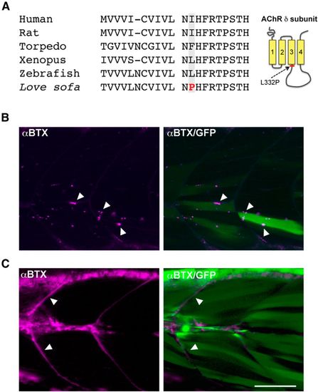
Genetic identification of the love sofa mutation. A, Alignment of amino acid sequences of AChR δ subunit from human, rat, torpedo, Xenopus, wild-type zebrafish, and the love sofa mutant. The putative mutation of love sofa is located at the shadowed amino acid. In wild-type zebrafish, the amino acid is leucine, whereas in other species isoleucine or phenylalanine is found. In love sofa, it is changed to proline. The location of the L332P mutation in the δ subunit protein is shown in the right panel. B, The wild-type δ subunit was expressed in mosaic fashion by injecting the gene construct into the love sofa mutant at one cell stage. Muscle fibers expressing the transgene, as marked by the cytoplasmic GFP (green), displayed distributed synapses (arrowheads) as marked by α-BTX (magenta). C, When the δ subunit harboring the L332P mutation was expressed in the sofa potato mutant, α-BTX staining visualized myoseptal synapses (arrowheads) but failed to exhibit distributed synapses. Magenta represents α-BTX; green represents cytoplasmic GFP. Scale bar, 50 μm. |
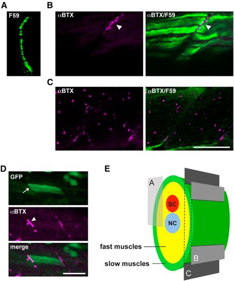
Different distribution of AChRs in slow and fast muscle fibers. A, Cross section of a wild-type trunk stained with the F59 antibody. The displayed region is indicated in E. F59-positive slow muscle cells form a single layer at the most superficial region. B, C, Optical slices of a wild-type larva near the surface (B) or deeper (C). Magenta represents α-BTX staining; green represents the F59 antibody signal. Scale bar, 50 μm. D, Stochastic expression of wild-type δ subunit in the sofa potato mutant. A slow muscle that expresses the transgene displayed cytoplasmic GFP (arrow; green) as well as α-BTX-positive AChR (arrowhead; magenta). Because fast muscles in the neighboring layer lacked the transgene expression, only myoseptal α-BTX staining was observed. Scale bar, 50 μm. E, Schematic cross section of the trunk of a larval zebrafish. Slow muscles are marked with green. Optical sections corresponding to A-C are indicated with a box and planes. SC, Spinal cord; NC, notochord. EXPRESSION / LABELING:
|
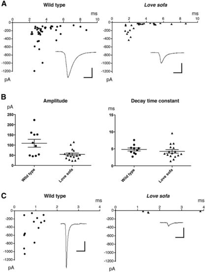
AChR functions in slow and fast fibers of the love sofa mutant. A, Decay time constants (x) plotted against current peak amplitudes (y) for mEPCs in a slow muscle of wild-type (left) or love sofa (right). Averaged traces for all mEPCs are shown as insets. Calibration: 100 pA, 5 ms. B, Accumulated data of current amplitudes and decay time constants recorded from slow muscle cells. Each dot represents an average for a single cell. Averages with SEs are shown. C, Decay time constants (x) plotted against current peak amplitudes (y) for mEPCs in fast muscle cells of wild-type (left) and love sofa (right). Averaged traces are shown as insets. Calibration: 100 pA, 5 ms. PHENOTYPE:
|

Nonfunctional AChRs in fast muscles of the love sofa mutant. A, Wild-type (left) and love sofa (right) larvae were doubly labeled with mAb35 antibody (green) and SV2 antibody (magenta). Boxes of dashed lines indicate myoseptal synapses in slow muscles. Arrowheads indicate distributed synapses in fast muscles. Scale bar, 50 μm. B, Representative traces of voltage-clamped slow and fast muscles in response to the application of 30 μM ACh. Calibration: 5 s, 500 pA. C, Amplitudes of ACh-induced currents in slow (n = 7) and fast muscles (n = 6) are shown. Each dot represents a muscle cell. EXPRESSION / LABELING:
PHENOTYPE:
|
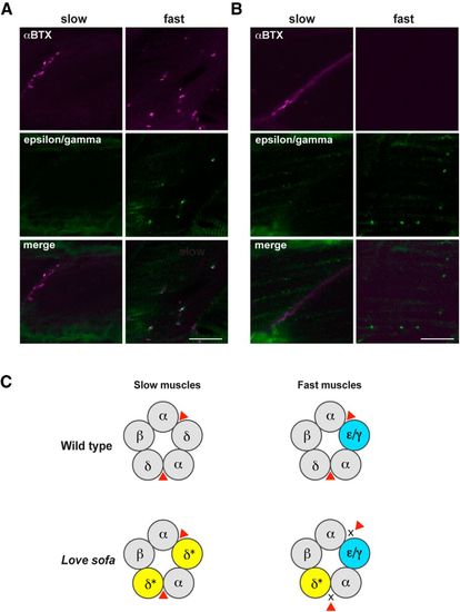
Distinct subunit compositions of AChRs in slow and fast muscles underlie the fiber-type specific phenotypes in the love sofa mutant. Wild-type (A) and love sofa (B) larvae were doubly labeled with an antibody for ε/γsubunits (green) and α-BTX (magenta). Optical sections corresponding to slow muscle fibers and fast muscle fibers are shown. Scale bar, 20 μm. C, A schematic depicting the hypothesis. In wild-type larvae (top), pentamers of slow muscles comprise αβδ, whereas those in fast muscle comprise αβδε/γ. Red triangle represents the ligand (ACh or α-BTX). In the love sofa mutant (bottom), the δ subunit harbors a L332P mutation (shown with an asterisk and yellow). As a result, the binding of ligand to the pockets between α and δ is inhibited in fast muscles. EXPRESSION / LABELING:
PHENOTYPE:
|