
Fig. 3
- ID
- ZDB-IMAGE-141112-3
- Genes
- Antibodies
- Publication
- Park et al., 2014 - A Single Mutation in the Acetylcholine Receptor δ-Subunit Causes Distinct Effects in Two Types of Neuromuscular Synapses
- All Figures
- Figures for Park et al., 2014
|
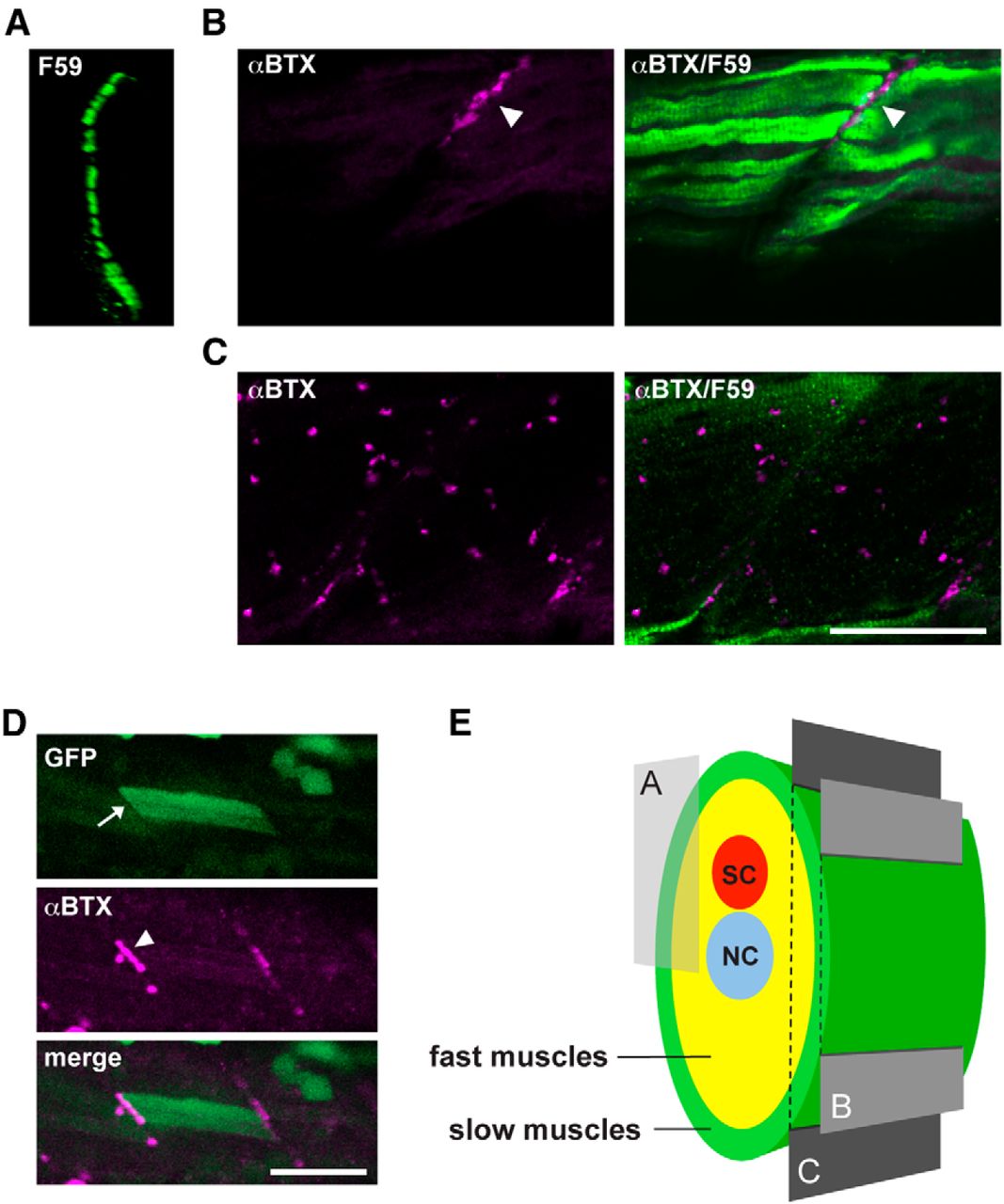
Fig. 3 Different distribution of AChRs in slow and fast muscle fibers. A, Cross section of a wild-type trunk stained with the F59 antibody. The displayed region is indicated in E. F59-positive slow muscle cells form a single layer at the most superficial region. B, C, Optical slices of a wild-type larva near the surface (B) or deeper (C). Magenta represents α-BTX staining; green represents the F59 antibody signal. Scale bar, 50 μm. D, Stochastic expression of wild-type δ subunit in the sofa potato mutant. A slow muscle that expresses the transgene displayed cytoplasmic GFP (arrow; green) as well as α-BTX-positive AChR (arrowhead; magenta). Because fast muscles in the neighboring layer lacked the transgene expression, only myoseptal α-BTX staining was observed. Scale bar, 50 μm. E, Schematic cross section of the trunk of a larval zebrafish. Slow muscles are marked with green. Optical sections corresponding to A-C are indicated with a box and planes. SC, Spinal cord; NC, notochord.