
- Title
-
Heart on a plate: histological and functional assessment of isolated adult zebrafish hearts maintained in culture
- Authors
- Pieperhoff, S., Wilson, K.S., Baily, J., de Mora, K., Maqsood, S., Vass, S., Taylor, J., Del-Pozo, J., MacRae, C.A., Mullins, J.J., Denvir, M.A.
- Source
- Full text @ PLoS One
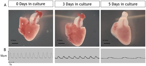
Low power images of excised cultured hearts (days 0–5). Upper panel (A) showing examples of excised hearts maintained in culture for 0, 3 and 5 days. Lower panel (B) shows example traces from Video-edge detection method displaying movement of the ventricle wall which was used to assess heart rate and ventricle function at days 0,3 and 5 in culture. |
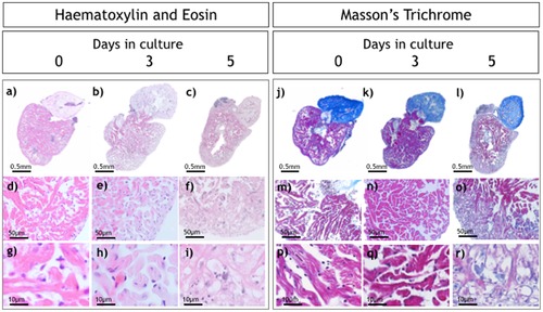
Histological analysis of isolated hearts. Haematoxylin and Eosin (H&E; a–i) and Masson’s trichrome staining (j–r) of hearts at 0, 3 and 5 days in culture. Higher magnification images (g, h, i) show normal cellular architecture at day 0 and day 3 with significant changes at day 5. |
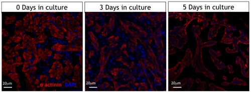
Immunohistology of isolated cultured hearts. Alpha-actinin immunostaining showing well preserved sarcomere patterns in cardiomyocytes of cultured hearts at day 0 and day 3 with loss of this typical pattern by day 5 in culture. (red is sarcomeric alpha-actinin (mouse, clone EA-53, Abcam), blue is DAPI (Serva Heidelberg)). |
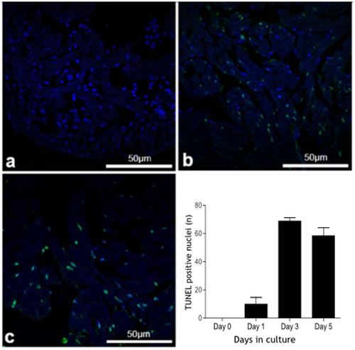
Apoptosis in isolated cultured zebrafish hearts (TUNEL staining). Apoptotic nuclei (green) and nuclear DAPI staining (blue) in zebrafish hearts (assessed by TUNEL staining) maintained in culture for 0, 1, 3 and 5 days showing a marked increase in the number of apoptotic bodies at day 3 which is maintained at day 5. |